Fabrication of GelMA – Agarose Based 3D Bioprinted Photocurable Hydrogel with In Vitro Cytocompatibility and Cells Mirroring Natural Keratocytes for Corneal Stromal Regeneration
Renuka Vijayaraghavan, Sravanthi Loganathan, Ravi Babu Valapa
{"title":"Fabrication of GelMA – Agarose Based 3D Bioprinted Photocurable Hydrogel with In Vitro Cytocompatibility and Cells Mirroring Natural Keratocytes for Corneal Stromal Regeneration","authors":"Renuka Vijayaraghavan, Sravanthi Loganathan, Ravi Babu Valapa","doi":"10.1002/mabi.202400136","DOIUrl":null,"url":null,"abstract":"<p>The complex anatomy of the cornea and the subsequent keratocyte-fibroblast transition have always made corneal stromal regeneration difficult. Recently, 3D printing has received considerable attention in terms of fabrication of scaffolds with precise dimension and pattern. In the current work, 3D printable polymer hydrogels made of GelMA/agarose are formulated and its rheological properties are evaluated. Despite the variation in agarose content, both the hydrogels exhibited G′>G′′ modulus. A prototype for 3D stromal model is created using Solid Works software, mimicking the anatomy of an adult cornea. The fabrication of 3D-printed hydrogels is performed using pneumatic extrusion. The FTIR analysis speculated that the hydrogel is well crosslinked and established strong hydrogen bonding with each other, thus contributing to improved thermal and structural stability. The MTT analysis revealed a higher rate of cell proliferation on the hydrogels. The optical analysis carried out on the 14th day of incubation revealed that the hydrogels exhibit transparency matching with natural corneal stromal tissue. Specific protein marker expression confirmed the keratocyte phenotype and showed that the cells do not undergo terminal differentiation into stromal fibroblasts. The findings of this work point to the potential of GelMA/A hydrogels as a novel biomaterial for corneal stromal tissue engineering.</p>","PeriodicalId":18103,"journal":{"name":"Macromolecular bioscience","volume":"24 10","pages":""},"PeriodicalIF":4.4000,"publicationDate":"2024-08-03","publicationTypes":"Journal Article","fieldsOfStudy":null,"isOpenAccess":false,"openAccessPdf":"","citationCount":"0","resultStr":null,"platform":"Semanticscholar","paperid":null,"PeriodicalName":"Macromolecular bioscience","FirstCategoryId":"5","ListUrlMain":"https://onlinelibrary.wiley.com/doi/10.1002/mabi.202400136","RegionNum":4,"RegionCategory":"医学","ArticlePicture":[],"TitleCN":null,"AbstractTextCN":null,"PMCID":null,"EPubDate":"","PubModel":"","JCR":"Q2","JCRName":"BIOCHEMISTRY & MOLECULAR BIOLOGY","Score":null,"Total":0}
引用次数: 0
Abstract
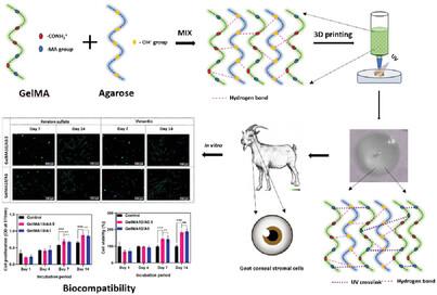
The complex anatomy of the cornea and the subsequent keratocyte-fibroblast transition have always made corneal stromal regeneration difficult. Recently, 3D printing has received considerable attention in terms of fabrication of scaffolds with precise dimension and pattern. In the current work, 3D printable polymer hydrogels made of GelMA/agarose are formulated and its rheological properties are evaluated. Despite the variation in agarose content, both the hydrogels exhibited G′>G′′ modulus. A prototype for 3D stromal model is created using Solid Works software, mimicking the anatomy of an adult cornea. The fabrication of 3D-printed hydrogels is performed using pneumatic extrusion. The FTIR analysis speculated that the hydrogel is well crosslinked and established strong hydrogen bonding with each other, thus contributing to improved thermal and structural stability. The MTT analysis revealed a higher rate of cell proliferation on the hydrogels. The optical analysis carried out on the 14th day of incubation revealed that the hydrogels exhibit transparency matching with natural corneal stromal tissue. Specific protein marker expression confirmed the keratocyte phenotype and showed that the cells do not undergo terminal differentiation into stromal fibroblasts. The findings of this work point to the potential of GelMA/A hydrogels as a novel biomaterial for corneal stromal tissue engineering.
期刊介绍:
Macromolecular Bioscience is a leading journal at the intersection of polymer and materials sciences with life science and medicine. With an Impact Factor of 2.895 (2018 Journal Impact Factor, Journal Citation Reports (Clarivate Analytics, 2019)), it is currently ranked among the top biomaterials and polymer journals.
Macromolecular Bioscience offers an attractive mixture of high-quality Reviews, Feature Articles, Communications, and Full Papers.
With average reviewing times below 30 days, publication times of 2.5 months and listing in all major indices, including Medline, Macromolecular Bioscience is the journal of choice for your best contributions at the intersection of polymer and life sciences.

 求助内容:
求助内容: 应助结果提醒方式:


