{"title":"Morphological and morphometric analysis of tarsal bones according to sex.","authors":"Nihal Gurlek Celik, Burcu Akman","doi":"10.1007/s00276-024-03450-6","DOIUrl":null,"url":null,"abstract":"<p><strong>Purpose: </strong>Our aim in the study is to measure the area and volume of the tarsal bones and examine the typing of the talus and calcaneus joint surfaces according to sex.</p><p><strong>Methods: </strong>In our study, the area and volume measurements of 630 tarsal bones and the morphology of the talus/calcaneus were analyzed by transferring thin-section Computed Tomography (CT) images to the 3D Slicer program.</p><p><strong>Results: </strong>The volume and area sizes of the foot bones are calcaneus, talus, cuboid, navicular, medial cuneiform, lateral cuneiform, and intermediate cuneiform, respectively. All area and volume values of males were statistically higher than females (p < 0.05). The right side calcaneus area, intermediate cuneiform area, and lateral cuneiform area values were statistically higher than the left side (p < 0.045, p < 0.044, p < 0.030, respectively). There was no statistical relationship between age and area/volume values (p > 0.05). Three different types were seen in the calcaneus and seven in the talus. The most common type in the calcaneus was B1 (40%), and the least common type was A (27.8%). Regardless of the subgroups, the most common type in the talus was type B (37.8%), while the least common type was E2 (1.1%).</p><p><strong>Conclusion: </strong>Although morphometric measurements of tarsal bones differed according to sex, they did not differ according to age. The frequency of occurrence of the types of articular surfaces of the talus and calcaneus varies according to populations. We think that the morphometry and morphology of tarsal bones will contribute to invasive procedures regarding tarsal bones and surrounding structures, and that three-dimensional bone modeling can be used to create educational materials.</p>","PeriodicalId":49461,"journal":{"name":"Surgical and Radiologic Anatomy","volume":null,"pages":null},"PeriodicalIF":1.4000,"publicationDate":"2024-08-02","publicationTypes":"Journal Article","fieldsOfStudy":null,"isOpenAccess":false,"openAccessPdf":"","citationCount":"0","resultStr":null,"platform":"Semanticscholar","paperid":null,"PeriodicalName":"Surgical and Radiologic Anatomy","FirstCategoryId":"3","ListUrlMain":"https://doi.org/10.1007/s00276-024-03450-6","RegionNum":4,"RegionCategory":"医学","ArticlePicture":[],"TitleCN":null,"AbstractTextCN":null,"PMCID":null,"EPubDate":"","PubModel":"","JCR":"Q2","JCRName":"Medicine","Score":null,"Total":0}
引用次数: 0
Abstract
Purpose: Our aim in the study is to measure the area and volume of the tarsal bones and examine the typing of the talus and calcaneus joint surfaces according to sex.
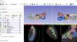
Methods: In our study, the area and volume measurements of 630 tarsal bones and the morphology of the talus/calcaneus were analyzed by transferring thin-section Computed Tomography (CT) images to the 3D Slicer program.
Results: The volume and area sizes of the foot bones are calcaneus, talus, cuboid, navicular, medial cuneiform, lateral cuneiform, and intermediate cuneiform, respectively. All area and volume values of males were statistically higher than females (p < 0.05). The right side calcaneus area, intermediate cuneiform area, and lateral cuneiform area values were statistically higher than the left side (p < 0.045, p < 0.044, p < 0.030, respectively). There was no statistical relationship between age and area/volume values (p > 0.05). Three different types were seen in the calcaneus and seven in the talus. The most common type in the calcaneus was B1 (40%), and the least common type was A (27.8%). Regardless of the subgroups, the most common type in the talus was type B (37.8%), while the least common type was E2 (1.1%).
Conclusion: Although morphometric measurements of tarsal bones differed according to sex, they did not differ according to age. The frequency of occurrence of the types of articular surfaces of the talus and calcaneus varies according to populations. We think that the morphometry and morphology of tarsal bones will contribute to invasive procedures regarding tarsal bones and surrounding structures, and that three-dimensional bone modeling can be used to create educational materials.
期刊介绍:
Anatomy is a morphological science which cannot fail to interest the clinician. The practical application of anatomical research to clinical problems necessitates special adaptation and selectivity in choosing from numerous international works. Although there is a tendency to believe that meaningful advances in anatomy are unlikely, constant revision is necessary. Surgical and Radiologic Anatomy, the first international journal of Clinical anatomy has been created in this spirit.
Its goal is to serve clinicians, regardless of speciality-physicians, surgeons, radiologists or other specialists-as an indispensable aid with which they can improve their knowledge of anatomy. Each issue includes: Original papers, review articles, articles on the anatomical bases of medical, surgical and radiological techniques, articles of normal radiologic anatomy, brief reviews of anatomical publications of clinical interest.
Particular attention is given to high quality illustrations, which are indispensable for a better understanding of anatomical problems.
Surgical and Radiologic Anatomy is a journal written by anatomists for clinicians with a special interest in anatomy.

 求助内容:
求助内容: 应助结果提醒方式:


