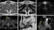
{"title":"IgG4-related disease with subcutaneous involvement and the associated diagnostic challenges with MRI.","authors":"Tomonori Kawasaki, Jiro Ichikawa, Kojiro Onohara, Satoshi Kanno, Masanori Wako, Naofumi Taniguchi, Satoshi Ochiai, Tomoaki Torigoe, Yasuo Yazawa","doi":"10.1007/s00256-024-04768-3","DOIUrl":null,"url":null,"abstract":"<p><p>IgG4-related disease is a rare fibroinflammatory disorder characterized by the infiltration of IgG4-rich plasma cells. Herein, we report a case of IgG4-related disease of the subcutaneous tissue with atypical MRI findings and difficulties in the histopathological examination using needle biopsy. Based on the clinical presentation and MRI findings, the patient was diagnosed with a benign myxoid or cystic tumor. Additionally, histopathological findings from a needle biopsy suggested a myxoma. Therefore, the correct diagnosis of IgG4-related disease was not made preoperatively. The resected specimens confirmed IgG4-related disease with an IgG4/IgG ratio > 80%. Previous reports have shown that the MRI findings of IgG4-related disease mimic both malignancy and inflammation; surprisingly, the features of subcutaneous IgG-related disease, including tail sign, unclear border, and heterogeneous enhancement, were similar to those found in sarcoma. Therefore, histopathological findings are needed for a correct diagnosis. Furthermore, careful examination is essential because the neoplasm and inflammation may overlap with IgG4-related disease, and needle biopsy is not fully reflective of the tumor. As is highlighted in the present case, IgG4-related disease is often misdiagnosed; therefore, clinicians should adequately recognize that even if the histopathological findings in biopsy were consistent with those observed in the MRI, misdiagnosis may occur.</p>","PeriodicalId":21783,"journal":{"name":"Skeletal Radiology","volume":" ","pages":"1147-1151"},"PeriodicalIF":1.9000,"publicationDate":"2025-05-01","publicationTypes":"Journal Article","fieldsOfStudy":null,"isOpenAccess":false,"openAccessPdf":"https://www.ncbi.nlm.nih.gov/pmc/articles/PMC11953168/pdf/","citationCount":"0","resultStr":null,"platform":"Semanticscholar","paperid":null,"PeriodicalName":"Skeletal Radiology","FirstCategoryId":"3","ListUrlMain":"https://doi.org/10.1007/s00256-024-04768-3","RegionNum":3,"RegionCategory":"医学","ArticlePicture":[],"TitleCN":null,"AbstractTextCN":null,"PMCID":null,"EPubDate":"2024/7/31 0:00:00","PubModel":"Epub","JCR":"Q2","JCRName":"ORTHOPEDICS","Score":null,"Total":0}
引用次数: 0
Abstract
IgG4-related disease is a rare fibroinflammatory disorder characterized by the infiltration of IgG4-rich plasma cells. Herein, we report a case of IgG4-related disease of the subcutaneous tissue with atypical MRI findings and difficulties in the histopathological examination using needle biopsy. Based on the clinical presentation and MRI findings, the patient was diagnosed with a benign myxoid or cystic tumor. Additionally, histopathological findings from a needle biopsy suggested a myxoma. Therefore, the correct diagnosis of IgG4-related disease was not made preoperatively. The resected specimens confirmed IgG4-related disease with an IgG4/IgG ratio > 80%. Previous reports have shown that the MRI findings of IgG4-related disease mimic both malignancy and inflammation; surprisingly, the features of subcutaneous IgG-related disease, including tail sign, unclear border, and heterogeneous enhancement, were similar to those found in sarcoma. Therefore, histopathological findings are needed for a correct diagnosis. Furthermore, careful examination is essential because the neoplasm and inflammation may overlap with IgG4-related disease, and needle biopsy is not fully reflective of the tumor. As is highlighted in the present case, IgG4-related disease is often misdiagnosed; therefore, clinicians should adequately recognize that even if the histopathological findings in biopsy were consistent with those observed in the MRI, misdiagnosis may occur.
期刊介绍:
Skeletal Radiology provides a forum for the dissemination of current knowledge and information dealing with disorders of the musculoskeletal system including the spine. While emphasizing the radiological aspects of the many varied skeletal abnormalities, the journal also adopts an interdisciplinary approach, reflecting the membership of the International Skeletal Society. Thus, the anatomical, pathological, physiological, clinical, metabolic and epidemiological aspects of the many entities affecting the skeleton receive appropriate consideration.
This is the Journal of the International Skeletal Society and the Official Journal of the Society of Skeletal Radiology and the Australasian Musculoskelelal Imaging Group.

 求助内容:
求助内容: 应助结果提醒方式:


