Mark J Kransdorf, Brandon T Larsen, Michael G Fox, Mark D Murphey, Jeremiah R Long
{"title":"Musculoskeletal glomus tumor: a review of 218 lesions in 176 patients.","authors":"Mark J Kransdorf, Brandon T Larsen, Michael G Fox, Mark D Murphey, Jeremiah R Long","doi":"10.1007/s00256-024-04743-y","DOIUrl":null,"url":null,"abstract":"<p><strong>Objective: </strong>To review the spectrum of clinical and imaging features of glomus tumor involving the musculoskeletal system including the typically solitary forms as well as the rarer multifocal forms (glomuvenous malformation and glomangiomatosis).</p><p><strong>Materials and methods: </strong>A retrospective review of our institutional pathology database from 1996 to 2023 identified 176 patients with 218 confirmed glomus tumors. Primary imaging studies included MRI (125), radiographs (100), clinical/intraoperative photos (77), and ultrasound (36). Lesions were divided into two groups: those that are typically solitary involving specific anatomic areas (finger, toe, soft tissue, coccyx, and bone), and those that are multifocal (glomuvenous malformation and glomangiomatosis).</p><p><strong>Results: </strong>The finger was the most frequently involved anatomic location for the classic (sporadic) glomus tumor occurring in 51% of patients, 77% of which were women, with the nail plate involved in more of the 75% of cases. Sporadic lesions involving the skin, subcutaneous adipose tissue, and deep soft tissue were termed \"soft tissue,\" and were identified in 39% of patients, 90% of which were in the extremities and in men in 81% of cases. The multifocal syndromic forms of glomus disease occurred in younger individuals and involved less than 6% of the study group. Patients with glomuvenous malformation presented early with predominantly cutaneous involvement, while those with glomangiomatosis present later, often with both superficial and deep involvement, and a high rate of local tumor recurrence.</p><p><strong>Conclusion: </strong>While glomus tumor is generally uncommon, it frequently involves the musculoskeletal extremities. Knowledge of the spectrum of characteristic locations and appearances will facilitate definitive diagnosis.</p>","PeriodicalId":21783,"journal":{"name":"Skeletal Radiology","volume":" ","pages":"457-479"},"PeriodicalIF":1.9000,"publicationDate":"2025-03-01","publicationTypes":"Journal Article","fieldsOfStudy":null,"isOpenAccess":false,"openAccessPdf":"","citationCount":"0","resultStr":null,"platform":"Semanticscholar","paperid":null,"PeriodicalName":"Skeletal Radiology","FirstCategoryId":"3","ListUrlMain":"https://doi.org/10.1007/s00256-024-04743-y","RegionNum":3,"RegionCategory":"医学","ArticlePicture":[],"TitleCN":null,"AbstractTextCN":null,"PMCID":null,"EPubDate":"2024/7/30 0:00:00","PubModel":"Epub","JCR":"Q2","JCRName":"ORTHOPEDICS","Score":null,"Total":0}
引用次数: 0
Abstract
Objective: To review the spectrum of clinical and imaging features of glomus tumor involving the musculoskeletal system including the typically solitary forms as well as the rarer multifocal forms (glomuvenous malformation and glomangiomatosis).
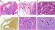
Materials and methods: A retrospective review of our institutional pathology database from 1996 to 2023 identified 176 patients with 218 confirmed glomus tumors. Primary imaging studies included MRI (125), radiographs (100), clinical/intraoperative photos (77), and ultrasound (36). Lesions were divided into two groups: those that are typically solitary involving specific anatomic areas (finger, toe, soft tissue, coccyx, and bone), and those that are multifocal (glomuvenous malformation and glomangiomatosis).
Results: The finger was the most frequently involved anatomic location for the classic (sporadic) glomus tumor occurring in 51% of patients, 77% of which were women, with the nail plate involved in more of the 75% of cases. Sporadic lesions involving the skin, subcutaneous adipose tissue, and deep soft tissue were termed "soft tissue," and were identified in 39% of patients, 90% of which were in the extremities and in men in 81% of cases. The multifocal syndromic forms of glomus disease occurred in younger individuals and involved less than 6% of the study group. Patients with glomuvenous malformation presented early with predominantly cutaneous involvement, while those with glomangiomatosis present later, often with both superficial and deep involvement, and a high rate of local tumor recurrence.
Conclusion: While glomus tumor is generally uncommon, it frequently involves the musculoskeletal extremities. Knowledge of the spectrum of characteristic locations and appearances will facilitate definitive diagnosis.
期刊介绍:
Skeletal Radiology provides a forum for the dissemination of current knowledge and information dealing with disorders of the musculoskeletal system including the spine. While emphasizing the radiological aspects of the many varied skeletal abnormalities, the journal also adopts an interdisciplinary approach, reflecting the membership of the International Skeletal Society. Thus, the anatomical, pathological, physiological, clinical, metabolic and epidemiological aspects of the many entities affecting the skeleton receive appropriate consideration.
This is the Journal of the International Skeletal Society and the Official Journal of the Society of Skeletal Radiology and the Australasian Musculoskelelal Imaging Group.

 求助内容:
求助内容: 应助结果提醒方式:


