{"title":"Clinical and pathological analyses of 14 cases of angiomatoid fibrous histiocytoma.","authors":"Qiang Zeng, Jie-Zhen Li, Guo-Ping Li, Yu-Peng Chen, Fang-Ling Song, Feng Gao","doi":"10.1007/s00795-024-00400-4","DOIUrl":null,"url":null,"abstract":"<p><p>Angiomatoid fibrous histiocytoma (AFH) is a soft tissue tumor of uncertain differentiation. Although its prognosis is good, its diagnosis and differential diagnosis remain a challenge, particularly for tumors with an atypical morphology. We evaluated the clinicopathological characteristics of 14 AFH cases and examined the key factors in its diagnosis or differential diagnosis. The cohort comprised 6 men and 8 women aged 9-65 years (average age: 31.2 years). Most of the tumors (11/14, 79%) were located in soft tissues, whereas 3/14 (21%) were located in the lung (1 case) and brain (2 cases). Tumor cells were spindle-shaped to epithelioid, with a visible fibrous capsule (9/14, 64%), hemorrhagic gap (9/14, 64%), lymphocyte sleeve (7/14, 50%), necrosis (3/14, 21%), and infiltrative boundary (4/14, 29%). The tumors expressed desmin (10/14, 71%) and exhibited low levels of Ki-67. 13 cases (93%) displayed ESWSR1 gene rearrangement. At follow-up, 1 case (7%) experienced local tumor recurrence. AFH is a rare intermediate tumor. Its pathological diagnosis requires a comprehensive analysis of histological, immunophenotypic, and molecular genetic features to avoid misdiagnosis. Our study has further enriched the histological features of AFH, emphasizing the importance of differential diagnosis and providing a reference for clinical practice.</p>","PeriodicalId":18338,"journal":{"name":"Medical Molecular Morphology","volume":" ","pages":"299-305"},"PeriodicalIF":1.1000,"publicationDate":"2024-12-01","publicationTypes":"Journal Article","fieldsOfStudy":null,"isOpenAccess":false,"openAccessPdf":"","citationCount":"0","resultStr":null,"platform":"Semanticscholar","paperid":null,"PeriodicalName":"Medical Molecular Morphology","FirstCategoryId":"3","ListUrlMain":"https://doi.org/10.1007/s00795-024-00400-4","RegionNum":4,"RegionCategory":"医学","ArticlePicture":[],"TitleCN":null,"AbstractTextCN":null,"PMCID":null,"EPubDate":"2024/7/30 0:00:00","PubModel":"Epub","JCR":"Q3","JCRName":"PATHOLOGY","Score":null,"Total":0}
引用次数: 0
Abstract
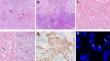
Angiomatoid fibrous histiocytoma (AFH) is a soft tissue tumor of uncertain differentiation. Although its prognosis is good, its diagnosis and differential diagnosis remain a challenge, particularly for tumors with an atypical morphology. We evaluated the clinicopathological characteristics of 14 AFH cases and examined the key factors in its diagnosis or differential diagnosis. The cohort comprised 6 men and 8 women aged 9-65 years (average age: 31.2 years). Most of the tumors (11/14, 79%) were located in soft tissues, whereas 3/14 (21%) were located in the lung (1 case) and brain (2 cases). Tumor cells were spindle-shaped to epithelioid, with a visible fibrous capsule (9/14, 64%), hemorrhagic gap (9/14, 64%), lymphocyte sleeve (7/14, 50%), necrosis (3/14, 21%), and infiltrative boundary (4/14, 29%). The tumors expressed desmin (10/14, 71%) and exhibited low levels of Ki-67. 13 cases (93%) displayed ESWSR1 gene rearrangement. At follow-up, 1 case (7%) experienced local tumor recurrence. AFH is a rare intermediate tumor. Its pathological diagnosis requires a comprehensive analysis of histological, immunophenotypic, and molecular genetic features to avoid misdiagnosis. Our study has further enriched the histological features of AFH, emphasizing the importance of differential diagnosis and providing a reference for clinical practice.
期刊介绍:
Medical Molecular Morphology is an international forum for researchers in both basic and clinical medicine to present and discuss new research on the structural mechanisms and the processes of health and disease at the molecular level. The structures of molecules, organelles, cells, tissues, and organs determine their normal function. Disease is thus best understood in terms of structural changes in these different levels of biological organization, especially in molecules and molecular interactions as well as the cellular localization of chemical components. Medical Molecular Morphology welcomes articles on basic or clinical research in the fields of cell biology, molecular biology, and medical, veterinary, and dental sciences using techniques for structural research such as electron microscopy, confocal laser scanning microscopy, enzyme histochemistry, immunohistochemistry, radioautography, X-ray microanalysis, and in situ hybridization.
Manuscripts submitted for publication must contain a statement to the effect that all human studies have been reviewed by the appropriate ethics committee and have therefore been performed in accordance with the ethical standards laid down in an appropriate version of the 1964 Declaration of Helsinki. It should also be stated clearly in the text that all persons gave their informed consent prior to their inclusion in the study. Details that might disclose the identity of the subjects under study should be omitted.
 求助内容:
求助内容: 应助结果提醒方式:


