Free-breathing 3D phase-resolved functional lung MRI vs breath-hold hyperpolarized 129Xe ventilation MRI in patients with chronic obstructive pulmonary disease and healthy volunteers.
IF 4.7 2区 医学Q1 RADIOLOGY, NUCLEAR MEDICINE & MEDICAL IMAGING
Filip Klimeš, Agilo Luitger Kern, Andreas Voskrebenzev, Marcel Gutberlet, Robert Grimm, Robin Aaron Müller, Lea Behrendt, Till Frederik Kaireit, Julian Glandorf, Tawfik Moher Alsady, Frank Wacker, Jens M Hohlfeld, Jens Vogel-Claussen
{"title":"Free-breathing 3D phase-resolved functional lung MRI vs breath-hold hyperpolarized <sup>129</sup>Xe ventilation MRI in patients with chronic obstructive pulmonary disease and healthy volunteers.","authors":"Filip Klimeš, Agilo Luitger Kern, Andreas Voskrebenzev, Marcel Gutberlet, Robert Grimm, Robin Aaron Müller, Lea Behrendt, Till Frederik Kaireit, Julian Glandorf, Tawfik Moher Alsady, Frank Wacker, Jens M Hohlfeld, Jens Vogel-Claussen","doi":"10.1007/s00330-024-10893-3","DOIUrl":null,"url":null,"abstract":"<p><strong>Objectives: </strong>3D phase-resolved functional lung (PREFUL) MRI offers evaluation of pulmonary ventilation without inhalation of contrast agent. This study seeks to compare ventilation maps obtained from 3D PREFUL MRI with a direct ventilation measurement derived from <sup>129</sup>Xe MRI in both patients with chronic obstructive pulmonary disease (COPD) and healthy volunteers.</p><p><strong>Methods: </strong>Thirty-one patients with COPD and 12 healthy controls underwent free-breathing 3D PREFUL MRI and breath-hold <sup>129</sup>Xe MRI at 1.5 T. For both MRI techniques, ventilation defect (VD) maps were determined and respective ventilation defect percentage (VDP) values were computed. All parameters of both techniques were compared by Spearman correlation coefficient (r) and the differences between VDP values were quantified by Bland-Altman analysis and tested for significance using Wilcoxon signed-rank test. In a regional comparison of VD maps, spatial overlap and Sørensen-Dice coefficients of healthy and defect areas were computed.</p><p><strong>Results: </strong>On a global level, all 3D PREFUL VDP values correlated significantly to VDP measure derived by <sup>129</sup>Xe ventilation imaging (all r > 0.65; all p < 0.0001). <sup>129</sup>Xe VDP was significantly greater than 3D PREFUL derived VDP<sub>RVent</sub> (mean bias = 10.5%, p < 0.001) and VDP<sub>FVL-CM</sub> (mean bias = 11.3%, p < 0.0001) but not for VDP<sub>Combined</sub> (mean bias = 1.7%, p = 0.70). The total regional agreement of <sup>129</sup>Xe and 3D PREFUL VD maps ranged between 60% and 63%.</p><p><strong>Conclusions: </strong>Free-breathing 3D PREFUL MRI showed a strong correlation with breath-hold hyperpolarized <sup>129</sup>Xe MRI regarding the VDP values and modest differences in the detection of VDs on a regional level.</p><p><strong>Clinical relevance statement: </strong>3D PREFUL MRI correlated with <sup>129</sup>Xe MRI, unveiling regional differences in COPD defect identification. This proposes 3D PREFUL MRI as a ventilation mapping surrogate, eliminating the need for extra hardware or inhaled gases.</p><p><strong>Key points: </strong>Current non-invasive evaluation techniques for lung diseases have drawbacks; <sup>129</sup>Xe MRI is limited by cost and availability. 3D PREFUL MRI correlated with <sup>129</sup>Xe MRI, with regional differences in identifying COPD defects. 3D PREFUL MRI can provide ventilation mapping without the need for additional hardware or inhaled gases.</p>","PeriodicalId":12076,"journal":{"name":"European Radiology","volume":" ","pages":"943-956"},"PeriodicalIF":4.7000,"publicationDate":"2025-02-01","publicationTypes":"Journal Article","fieldsOfStudy":null,"isOpenAccess":false,"openAccessPdf":"https://www.ncbi.nlm.nih.gov/pmc/articles/PMC11782336/pdf/","citationCount":"0","resultStr":null,"platform":"Semanticscholar","paperid":null,"PeriodicalName":"European Radiology","FirstCategoryId":"3","ListUrlMain":"https://doi.org/10.1007/s00330-024-10893-3","RegionNum":2,"RegionCategory":"医学","ArticlePicture":[],"TitleCN":null,"AbstractTextCN":null,"PMCID":null,"EPubDate":"2024/7/26 0:00:00","PubModel":"Epub","JCR":"Q1","JCRName":"RADIOLOGY, NUCLEAR MEDICINE & MEDICAL IMAGING","Score":null,"Total":0}
引用次数: 0
Abstract
Objectives: 3D phase-resolved functional lung (PREFUL) MRI offers evaluation of pulmonary ventilation without inhalation of contrast agent. This study seeks to compare ventilation maps obtained from 3D PREFUL MRI with a direct ventilation measurement derived from 129Xe MRI in both patients with chronic obstructive pulmonary disease (COPD) and healthy volunteers.
Methods: Thirty-one patients with COPD and 12 healthy controls underwent free-breathing 3D PREFUL MRI and breath-hold 129Xe MRI at 1.5 T. For both MRI techniques, ventilation defect (VD) maps were determined and respective ventilation defect percentage (VDP) values were computed. All parameters of both techniques were compared by Spearman correlation coefficient (r) and the differences between VDP values were quantified by Bland-Altman analysis and tested for significance using Wilcoxon signed-rank test. In a regional comparison of VD maps, spatial overlap and Sørensen-Dice coefficients of healthy and defect areas were computed.
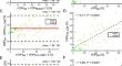
Results: On a global level, all 3D PREFUL VDP values correlated significantly to VDP measure derived by 129Xe ventilation imaging (all r > 0.65; all p < 0.0001). 129Xe VDP was significantly greater than 3D PREFUL derived VDPRVent (mean bias = 10.5%, p < 0.001) and VDPFVL-CM (mean bias = 11.3%, p < 0.0001) but not for VDPCombined (mean bias = 1.7%, p = 0.70). The total regional agreement of 129Xe and 3D PREFUL VD maps ranged between 60% and 63%.
Conclusions: Free-breathing 3D PREFUL MRI showed a strong correlation with breath-hold hyperpolarized 129Xe MRI regarding the VDP values and modest differences in the detection of VDs on a regional level.
Clinical relevance statement: 3D PREFUL MRI correlated with 129Xe MRI, unveiling regional differences in COPD defect identification. This proposes 3D PREFUL MRI as a ventilation mapping surrogate, eliminating the need for extra hardware or inhaled gases.
Key points: Current non-invasive evaluation techniques for lung diseases have drawbacks; 129Xe MRI is limited by cost and availability. 3D PREFUL MRI correlated with 129Xe MRI, with regional differences in identifying COPD defects. 3D PREFUL MRI can provide ventilation mapping without the need for additional hardware or inhaled gases.
期刊介绍:
European Radiology (ER) continuously updates scientific knowledge in radiology by publication of strong original articles and state-of-the-art reviews written by leading radiologists. A well balanced combination of review articles, original papers, short communications from European radiological congresses and information on society matters makes ER an indispensable source for current information in this field.
This is the Journal of the European Society of Radiology, and the official journal of a number of societies.
From 2004-2008 supplements to European Radiology were published under its companion, European Radiology Supplements, ISSN 1613-3749.

 求助内容:
求助内容: 应助结果提醒方式:


