Jessica Murphy, Abdulrahman Dera, José A. Morais, Michael A. Tsoukas, Natalie Khor, Taisiia Sazonova, Lucas Guimarães Almeida, Alexandra B. Cooke, Stella S. Daskalopoulou, Bjorn T. Tam, Sylvia Santosa
{"title":"Age of obesity onset affects subcutaneous adipose tissue cellularity differently in the abdominal and femoral region","authors":"Jessica Murphy, Abdulrahman Dera, José A. Morais, Michael A. Tsoukas, Natalie Khor, Taisiia Sazonova, Lucas Guimarães Almeida, Alexandra B. Cooke, Stella S. Daskalopoulou, Bjorn T. Tam, Sylvia Santosa","doi":"10.1002/oby.24059","DOIUrl":null,"url":null,"abstract":"<div>\n \n \n <section>\n \n <h3> Objective</h3>\n \n <p>We aimed to examine the effect of age of obesity onset, sex, and their interaction on abdominal and femoral subcutaneous adipose tissue (SAT) morphology (degree of adipocyte hyperplasia or hypertrophy).</p>\n </section>\n \n <section>\n \n <h3> Methods</h3>\n \n <p>In this cross-sectional study, we isolated adipocytes via collagenase digestion from abdominal and femoral SAT biopsies taken from male and female adults with childhood-onset obesity (CO; <i>n</i> = 8 males, <i>n</i> = 16 females) or adult-onset obesity (AO; <i>n</i> = 8 males, <i>n</i> = 13 females). Regional body composition was measured with dual-energy x-ray absorptiometry and a single-slice abdominal computed tomography scan. Mean adipocyte size was measured in abdominal and femoral SAT and was used to quantify morphology in android and gynoid subcutaneous fat, respectively.</p>\n </section>\n \n <section>\n \n <h3> Results</h3>\n \n <p>Abdominal SAT morphology was more hyperplastic in females with CO than females with AO (<i>p</i> = 0.004) but did not differ between males with CO and males with AO (<i>p</i> = 0.996). Conversely, femoral SAT morphology was more hypertrophic in males and females with CO than those with AO.</p>\n </section>\n \n <section>\n \n <h3> Conclusions</h3>\n \n <p>Age of obesity onset appears to affect SAT morphology differently in the abdominal and femoral regions of male and female adults. Our findings challenge the notion that SAT is uniformly hyperplastic in CO and hypertrophic in AO.</p>\n </section>\n </div>","PeriodicalId":215,"journal":{"name":"Obesity","volume":"32 8","pages":"1508-1517"},"PeriodicalIF":4.2000,"publicationDate":"2024-07-24","publicationTypes":"Journal Article","fieldsOfStudy":null,"isOpenAccess":false,"openAccessPdf":"https://onlinelibrary.wiley.com/doi/epdf/10.1002/oby.24059","citationCount":"0","resultStr":null,"platform":"Semanticscholar","paperid":null,"PeriodicalName":"Obesity","FirstCategoryId":"3","ListUrlMain":"https://onlinelibrary.wiley.com/doi/10.1002/oby.24059","RegionNum":2,"RegionCategory":"医学","ArticlePicture":[],"TitleCN":null,"AbstractTextCN":null,"PMCID":null,"EPubDate":"","PubModel":"","JCR":"Q1","JCRName":"ENDOCRINOLOGY & METABOLISM","Score":null,"Total":0}
引用次数: 0
Abstract
Objective
We aimed to examine the effect of age of obesity onset, sex, and their interaction on abdominal and femoral subcutaneous adipose tissue (SAT) morphology (degree of adipocyte hyperplasia or hypertrophy).
Methods
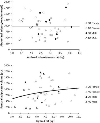
In this cross-sectional study, we isolated adipocytes via collagenase digestion from abdominal and femoral SAT biopsies taken from male and female adults with childhood-onset obesity (CO; n = 8 males, n = 16 females) or adult-onset obesity (AO; n = 8 males, n = 13 females). Regional body composition was measured with dual-energy x-ray absorptiometry and a single-slice abdominal computed tomography scan. Mean adipocyte size was measured in abdominal and femoral SAT and was used to quantify morphology in android and gynoid subcutaneous fat, respectively.
Results
Abdominal SAT morphology was more hyperplastic in females with CO than females with AO (p = 0.004) but did not differ between males with CO and males with AO (p = 0.996). Conversely, femoral SAT morphology was more hypertrophic in males and females with CO than those with AO.
Conclusions
Age of obesity onset appears to affect SAT morphology differently in the abdominal and femoral regions of male and female adults. Our findings challenge the notion that SAT is uniformly hyperplastic in CO and hypertrophic in AO.
研究目的我们旨在研究肥胖发病年龄、性别及其相互作用对腹部和股部皮下脂肪组织(SAT)形态(脂肪细胞增生或肥大程度)的影响:在这项横断面研究中,我们通过胶原酶消化从患有儿童期肥胖症(CO;男性8人,女性16人)或成年期肥胖症(AO;男性8人,女性13人)的男性和女性成人的腹部和股部SAT活检组织中分离出脂肪细胞。通过双能 X 射线吸收测量法和单片腹部计算机断层扫描测量区域身体成分。测量了腹部和股部SAT脂肪细胞的平均大小,并分别用于量化甲状腺和妇科皮下脂肪的形态:结果:患有 CO 的女性腹部 SAT 形态比患有 AO 的女性更加增生(p = 0.004),但患有 CO 的男性和患有 AO 的男性之间没有差异(p = 0.996)。相反,患有CO的男性和女性的股骨SAT形态比患有AO的男性和女性更肥大:结论:肥胖症的发病年龄似乎会对男性和女性成年人腹部和股部的SAT形态产生不同的影响。我们的研究结果对SAT在CO患者中均匀增生而在AO患者中肥大的观点提出了质疑。
期刊介绍:
Obesity is the official journal of The Obesity Society and is the premier source of information for increasing knowledge, fostering translational research from basic to population science, and promoting better treatment for people with obesity. Obesity publishes important peer-reviewed research and cutting-edge reviews, commentaries, and public health and medical developments.
 求助内容:
求助内容: 应助结果提醒方式:


