Clinicopathologic and molecular study of superficial CD34-positive fibroblastic tumours mimicking atypical fibrous histiocytoma (dermatofibroma).

IF 3.9 2区 医学 Q2 CELL BIOLOGY
Histopathology Pub Date : 2024-12-01 Epub Date: 2024-07-24 DOI:10.1111/his.15282
Craig B Wakefield, Fredrik Mertens, Christopher D M Fletcher, William J Anderson
{"title":"Clinicopathologic and molecular study of superficial CD34-positive fibroblastic tumours mimicking atypical fibrous histiocytoma (dermatofibroma).","authors":"Craig B Wakefield, Fredrik Mertens, Christopher D M Fletcher, William J Anderson","doi":"10.1111/his.15282","DOIUrl":null,"url":null,"abstract":"<p><strong>Aims: </strong>Superficial CD34-positive fibroblastic tumour (SCD34FT) is an uncommon but distinctive low-grade neoplasm of the skin and subcutis that shows frequent CADM3 expression by immunohistochemistry (IHC). In this study, prompted by an index case resembling 'atypical fibrous histiocytoma (FH)' that was positive for CADM3 IHC, we systematically examined a cohort of tumours previously diagnosed as 'atypical FH' by applying CADM3 and fluorescence in situ hybridization (FISH) for PRDM10 rearrangement, to investigate the overlap between these tumour types.</p><p><strong>Methods and results: </strong>Forty cases of atypical FH were retrieved, including CD34-positive tumours (n = 20) and CD34-negative tumours (n = 20). All tumours were stained for CADM3. All CADM3-positive tumours were evaluated by FISH to assess for PRDM10 rearrangement. Eleven CD34-positive tumours (11/20, 55%) coexpressed CADM3 and were reclassified as SCD34FT. None (0/20) of the CD34-negative atypical FH were CADM3-positive. Reclassified SCD34FT (10/11) arose on the lower extremity, with frequent involvement of the thigh (n = 8). Features suggestive of atypical FH were observed in many reclassified cases including variable cellularity, spindled morphology, infiltrative tumour margins, collagen entrapment, epidermal hyperpigmentation, and acanthosis. Variably prominent multinucleate giant cells, including Touton-like forms, were also present. An informative FISH result was obtained in 10/11 reclassified tumours, with 60% (6/10) demonstrating PRDM10 rearrangement.</p><p><strong>Conclusion: </strong>A significant subset of tumours that histologically resemble atypical FH, and are positive for CD34, coexpress CADM3 and harbour PRDM10 rearrangement, supporting their reclassification as SCD34FT. Awareness of this morphologic overlap and the application of CADM3 IHC can aid the distinction between SCD34FT and atypical FH.</p>","PeriodicalId":13219,"journal":{"name":"Histopathology","volume":null,"pages":null},"PeriodicalIF":3.9000,"publicationDate":"2024-12-01","publicationTypes":"Journal Article","fieldsOfStudy":null,"isOpenAccess":false,"openAccessPdf":"","citationCount":"0","resultStr":null,"platform":"Semanticscholar","paperid":null,"PeriodicalName":"Histopathology","FirstCategoryId":"3","ListUrlMain":"https://doi.org/10.1111/his.15282","RegionNum":2,"RegionCategory":"医学","ArticlePicture":[],"TitleCN":null,"AbstractTextCN":null,"PMCID":null,"EPubDate":"2024/7/24 0:00:00","PubModel":"Epub","JCR":"Q2","JCRName":"CELL BIOLOGY","Score":null,"Total":0}
引用次数: 0

Abstract

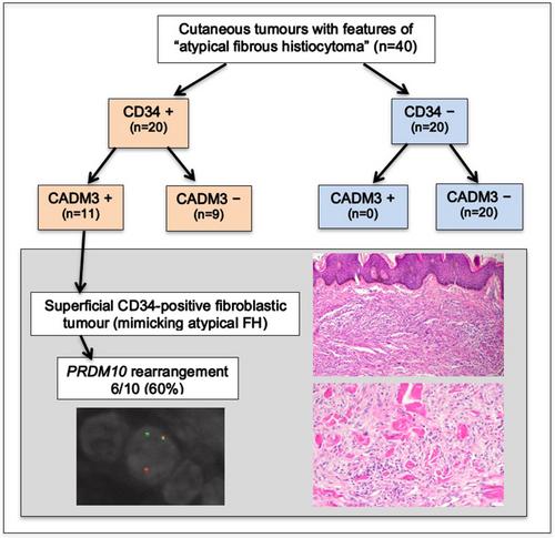
Aims: Superficial CD34-positive fibroblastic tumour (SCD34FT) is an uncommon but distinctive low-grade neoplasm of the skin and subcutis that shows frequent CADM3 expression by immunohistochemistry (IHC). In this study, prompted by an index case resembling 'atypical fibrous histiocytoma (FH)' that was positive for CADM3 IHC, we systematically examined a cohort of tumours previously diagnosed as 'atypical FH' by applying CADM3 and fluorescence in situ hybridization (FISH) for PRDM10 rearrangement, to investigate the overlap between these tumour types.

Methods and results: Forty cases of atypical FH were retrieved, including CD34-positive tumours (n = 20) and CD34-negative tumours (n = 20). All tumours were stained for CADM3. All CADM3-positive tumours were evaluated by FISH to assess for PRDM10 rearrangement. Eleven CD34-positive tumours (11/20, 55%) coexpressed CADM3 and were reclassified as SCD34FT. None (0/20) of the CD34-negative atypical FH were CADM3-positive. Reclassified SCD34FT (10/11) arose on the lower extremity, with frequent involvement of the thigh (n = 8). Features suggestive of atypical FH were observed in many reclassified cases including variable cellularity, spindled morphology, infiltrative tumour margins, collagen entrapment, epidermal hyperpigmentation, and acanthosis. Variably prominent multinucleate giant cells, including Touton-like forms, were also present. An informative FISH result was obtained in 10/11 reclassified tumours, with 60% (6/10) demonstrating PRDM10 rearrangement.

Conclusion: A significant subset of tumours that histologically resemble atypical FH, and are positive for CD34, coexpress CADM3 and harbour PRDM10 rearrangement, supporting their reclassification as SCD34FT. Awareness of this morphologic overlap and the application of CADM3 IHC can aid the distinction between SCD34FT and atypical FH.
模仿非典型纤维组织细胞瘤(皮肤纤维瘤)的浅表 CD34 阳性成纤维细胞瘤的临床病理和分子研究。
目的:表皮 CD34 阳性成纤维细胞瘤(SCD34FT)是皮肤和皮下的一种不常见但很独特的低级别肿瘤,免疫组化(IHC)显示其经常表达 CADM3。在本研究中,由于一个类似于 "非典型纤维组织细胞瘤(FH)"的病例在 CADM3 IHC 中呈阳性,我们通过应用 CADM3 和荧光原位杂交(FISH)检测 PRDM10 重排,系统地检查了一组以前被诊断为 "非典型 FH "的肿瘤,以研究这些肿瘤类型之间的重叠:检索了40例非典型FH,包括CD34阳性肿瘤(20例)和CD34阴性肿瘤(20例)。所有肿瘤均进行了 CADM3 染色。所有CADM3阳性肿瘤均通过FISH评估PRDM10重排。11个CD34阳性肿瘤(11/20,55%)共表达CADM3,被重新归类为SCD34FT。CD34 阴性的非典型 FH 中没有一个(0/20)CADM3 阳性。重新分类的 SCD34FT(10/11)出现在下肢,大腿经常受累(n = 8)。在许多重新分类的病例中都观察到了提示非典型 FH 的特征,包括可变细胞性、纺锤形形态、肿瘤边缘浸润、胶原夹层、表皮色素沉着和棘皮症。此外,还出现了不同程度的突出多核巨细胞,包括 Touton 样细胞。10/11例重新分类的肿瘤都获得了有参考价值的FISH结果,其中60%(6/10)的肿瘤显示了PRDM10重排:结论:有相当一部分肿瘤在组织学上与非典型 FH 相似,CD34 阳性,同时表达 CADM3 和携带 PRDM10 重排,支持将其重新分类为 SCD34FT。对这种形态学重叠的认识和 CADM3 IHC 的应用有助于区分 SCD34FT 和非典型 FH。
本文章由计算机程序翻译,如有差异,请以英文原文为准。
求助全文
约1分钟内获得全文 求助全文
来源期刊
Histopathology
Histopathology 医学-病理学
CiteScore
10.20
自引率
4.70%
发文量
239
审稿时长
1 months
期刊介绍: Histopathology is an international journal intended to be of practical value to surgical and diagnostic histopathologists, and to investigators of human disease who employ histopathological methods. Our primary purpose is to publish advances in pathology, in particular those applicable to clinical practice and contributing to the better understanding of human disease.
×
引用
GB/T 7714-2015
复制
MLA
复制
APA
复制
导出至
BibTeX EndNote RefMan NoteFirst NoteExpress
×
提示
您的信息不完整,为了账户安全,请先补充。
现在去补充
×
提示
您因"违规操作"
具体请查看互助需知
我知道了
×
提示
确定
请完成安全验证×
copy
已复制链接
快去分享给好友吧!
我知道了
右上角分享
点击右上角分享
0
联系我们:info@booksci.cn Book学术提供免费学术资源搜索服务,方便国内外学者检索中英文文献。致力于提供最便捷和优质的服务体验。 Copyright © 2023 布克学术 All rights reserved.
京ICP备2023020795号-1
ghs 京公网安备 11010802042870号
Book学术文献互助
Book学术文献互助群
群 号:481959085
Book学术官方微信