Renal denervation alleviates vascular remodeling in spontaneously hypertensive rats by regulating perivascular adipose tissue
IF 4.3
2区 医学
Q1 PERIPHERAL VASCULAR DISEASE
引用次数: 0
Abstract
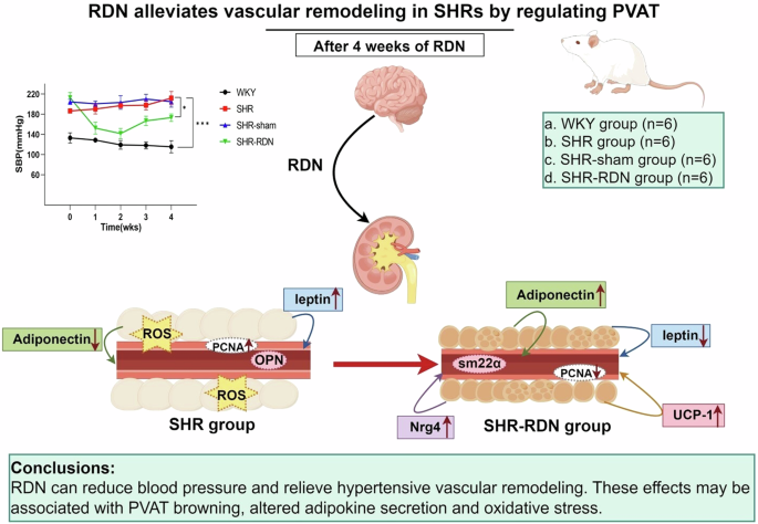
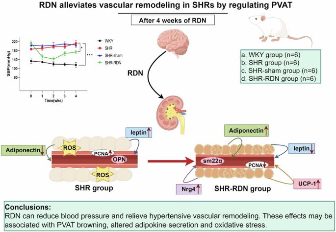
Vascular remodeling is the main pathological process that causes the damage of the target organ of hypertension. Perivascular adipose tissue (PVAT) surrounds blood vessels and plays a key role in the pathogenesis of various cardiovascular diseases. This study aimed to investigate the effects of renal denervation (RDN) on hypertensive vascular remodeling and to elucidate the role of PVAT in this process. Male spontaneously hypertensive rat (SHR) and Wistar-Kyoto (WKY) rat were selected. Aortic vascular remodeling was evaluated using hematoxylin and eosin (H&E) staining and Masson’s trichrome staining. Morphological changes in the PVAT were observed through H&E and Oil Red O staining. Dihydroethidium was used to measure oxidative stress levels in PVAT, while western blot analysis was used to determine the expression levels of proteins associated with vascular remodeling. The results showed that the aortic medial thickness, media thickness/lumen diameter, collagen volume fraction, and reactive oxygen species (ROS) level in PVAT were significantly higher in the SHR group than in the WKY group. The indexes mentioned above were lower in the SHR-RDN group than in the SHR group. H&E staining revealed that fat droplets in PVAT in the SHR-RDN group became smaller and browning occurred. Moreover, the protein expression of uncoupling protein-1 (UCP-1) and neuregulin 4 (Nrg4) was significantly increased in the SHR-RDN group. In addition, the expression of adiponectin increased and the expression of leptin decreased in the SHR-RDN group compared to the SHR group. In conclusion, RDN can relieve hypertensive vascular remodeling, which may be associated with PVAT.


肾脏去神经通过调节血管周围脂肪组织减轻自发性高血压大鼠的血管重塑。
血管重塑是导致高血压靶器官损伤的主要病理过程。血管周围脂肪组织(PVAT)包围着血管,在各种心血管疾病的发病机制中起着关键作用。本研究旨在探讨肾脏去神经(RDN)对高血压血管重塑的影响,并阐明 PVAT 在这一过程中的作用。研究选择了雄性自发性高血压大鼠(SHR)和 Wistar-Kyoto 大鼠(WKY)。使用苏木精和伊红(H&E)染色法和马森三色染色法评估主动脉血管重塑。通过 H&E 和油红 O 染色观察 PVAT 的形态变化。二氢乙锭用于测量 PVAT 的氧化应激水平,而 Western 印迹分析则用于确定与血管重塑相关的蛋白质的表达水平。结果显示,SHR 组主动脉内膜厚度、介质厚度/管腔直径、胶原体积分数和 PVAT 中活性氧(ROS)水平显著高于 WKY 组。SHR-RDN组的上述指标均低于SHR组。H&E染色显示,SHR-RDN组PVAT中的脂肪滴变小,并出现褐变。此外,解偶联蛋白-1(UCP-1)和神经胶质蛋白4(Nrg4)的蛋白表达在SHR-RDN组显著增加。此外,与 SHR 组相比,SHR-RDN 组脂肪连素的表达增加,瘦素的表达减少。总之,RDN能缓解高血压血管重塑,而血管重塑可能与PVAT有关。
本文章由计算机程序翻译,如有差异,请以英文原文为准。
求助全文
约1分钟内获得全文
求助全文
来源期刊

Hypertension Research
医学-外周血管病
CiteScore
7.40
自引率
16.70%
发文量
249
审稿时长
3-8 weeks
期刊介绍:
Hypertension Research is the official publication of the Japanese Society of Hypertension. The journal publishes papers reporting original clinical and experimental research that contribute to the advancement of knowledge in the field of hypertension and related cardiovascular diseases. The journal publishes Review Articles, Articles, Correspondence and Comments.
 求助内容:
求助内容: 应助结果提醒方式:


