Arianna Barbotti, Alexandru Szathmari, Matthieu Vinchon, Pierre-Aurélien Beuriat, Federico Di Rocco
{"title":"Piezosurgery in endoscopic-assisted trigonocephaly correction: a technical note.","authors":"Arianna Barbotti, Alexandru Szathmari, Matthieu Vinchon, Pierre-Aurélien Beuriat, Federico Di Rocco","doi":"10.1007/s00381-024-06551-0","DOIUrl":null,"url":null,"abstract":"<p><strong>Purpose: </strong>This study aims to evaluate the effectiveness of the Piezosurgery® device in endoscopic-assisted correction of trigonocephaly. Trigonocephaly is a type of craniosynostosis characterized by a triangular-shaped forehead due to the premature fusion of the metopic suture. Traditional open cranial vault reconstruction, although common, is invasive and poses risks. The study explores a less invasive alternative using ultrasonic microvibrations for bone cutting, potentially reducing soft tissue damage and improving surgical outcomes.</p><p><strong>Methods: </strong>The Piezosurgery® device was employed in endoscopic trigonocephaly correction surgeries performed on patients under 4 months old at the French Referral Center for Craniosynostosis in Lyon. The technique involves making a small skin incision and performing osteotomies from the anterior fontanel to the glabella. A rigid 0° endoscope provides visibility, and the Piezosurgery® device enables precise bone cutting while preserving the dura mater. Post-surgery, patients were discharged within 3 days and required to wear a remodeling helmet for 6-8 months.</p><p><strong>Results: </strong>The use of Piezosurgery® device allowed precise osteotomies with minimal soft tissue damage. No dura mater injuries occurred in the patient series. The procedure was efficient, with an average duration of 80 min, and blood loss was minimal, reducing the need for blood transfusions. The endoscopic approach facilitated shorter surgical times and reduced postoperative infection risks. Enhanced visibility during surgery, due to cavitation effects, improved the accuracy of bone cuts. The technique demonstrated promising safety and esthetic outcomes, although it incurred higher costs compared to traditional methods.</p><p><strong>Conclusion: </strong>Piezosurgery® device provides a safe and effective method for minimally invasive endoscopic correction of trigonocephaly. The device's ability to selectively cut bone while preserving soft tissues offers significant advantages, despite longer surgical times and higher costs. This technique represents a viable alternative to traditional open surgery, promoting better clinical outcomes and reduced recovery times.</p>","PeriodicalId":9970,"journal":{"name":"Child's Nervous System","volume":null,"pages":null},"PeriodicalIF":1.3000,"publicationDate":"2024-09-01","publicationTypes":"Journal Article","fieldsOfStudy":null,"isOpenAccess":false,"openAccessPdf":"","citationCount":"0","resultStr":null,"platform":"Semanticscholar","paperid":null,"PeriodicalName":"Child's Nervous System","FirstCategoryId":"3","ListUrlMain":"https://doi.org/10.1007/s00381-024-06551-0","RegionNum":4,"RegionCategory":"医学","ArticlePicture":[],"TitleCN":null,"AbstractTextCN":null,"PMCID":null,"EPubDate":"2024/7/23 0:00:00","PubModel":"Epub","JCR":"Q4","JCRName":"CLINICAL NEUROLOGY","Score":null,"Total":0}
引用次数: 0
Abstract
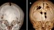
Purpose: This study aims to evaluate the effectiveness of the Piezosurgery® device in endoscopic-assisted correction of trigonocephaly. Trigonocephaly is a type of craniosynostosis characterized by a triangular-shaped forehead due to the premature fusion of the metopic suture. Traditional open cranial vault reconstruction, although common, is invasive and poses risks. The study explores a less invasive alternative using ultrasonic microvibrations for bone cutting, potentially reducing soft tissue damage and improving surgical outcomes.
Methods: The Piezosurgery® device was employed in endoscopic trigonocephaly correction surgeries performed on patients under 4 months old at the French Referral Center for Craniosynostosis in Lyon. The technique involves making a small skin incision and performing osteotomies from the anterior fontanel to the glabella. A rigid 0° endoscope provides visibility, and the Piezosurgery® device enables precise bone cutting while preserving the dura mater. Post-surgery, patients were discharged within 3 days and required to wear a remodeling helmet for 6-8 months.
Results: The use of Piezosurgery® device allowed precise osteotomies with minimal soft tissue damage. No dura mater injuries occurred in the patient series. The procedure was efficient, with an average duration of 80 min, and blood loss was minimal, reducing the need for blood transfusions. The endoscopic approach facilitated shorter surgical times and reduced postoperative infection risks. Enhanced visibility during surgery, due to cavitation effects, improved the accuracy of bone cuts. The technique demonstrated promising safety and esthetic outcomes, although it incurred higher costs compared to traditional methods.
Conclusion: Piezosurgery® device provides a safe and effective method for minimally invasive endoscopic correction of trigonocephaly. The device's ability to selectively cut bone while preserving soft tissues offers significant advantages, despite longer surgical times and higher costs. This technique represents a viable alternative to traditional open surgery, promoting better clinical outcomes and reduced recovery times.
期刊介绍:
The journal has been expanded to encompass all aspects of pediatric neurosciences concerning the developmental and acquired abnormalities of the nervous system and its coverings, functional disorders, epilepsy, spasticity, basic and clinical neuro-oncology, rehabilitation and trauma. Global pediatric neurosurgery is an additional field of interest that will be considered for publication in the journal.
 求助内容:
求助内容: 应助结果提醒方式:


