Combined matrix-assisted laser desorption/ionisation-mass spectrometry imaging with liquid chromatography-tandem mass spectrometry for observing spatial distribution of lipids in whole Caenorhabditis elegans

IF 1.8 3区 化学 Q4 BIOCHEMICAL RESEARCH METHODS
Michiel Vandenbosch, Erika R. Amstalden van Hove, Ronny Mohren, Isabeau Vermeulen, Henry Dijkman, Ron M. A. Heeren, Pim E. G. Leonards, Samantha Hughes
{"title":"Combined matrix-assisted laser desorption/ionisation-mass spectrometry imaging with liquid chromatography-tandem mass spectrometry for observing spatial distribution of lipids in whole Caenorhabditis elegans","authors":"Michiel Vandenbosch,&nbsp;Erika R. Amstalden van Hove,&nbsp;Ronny Mohren,&nbsp;Isabeau Vermeulen,&nbsp;Henry Dijkman,&nbsp;Ron M. A. Heeren,&nbsp;Pim E. G. Leonards,&nbsp;Samantha Hughes","doi":"10.1002/rcm.9850","DOIUrl":null,"url":null,"abstract":"<div>\n \n <section>\n \n <h3> Rationale</h3>\n \n <p>Matrix-assisted laser desorption/ionisation-mass spectrometry imaging (MALDI-MSI) is a powerful label-free technique for biomolecule detection (e.g., lipids), within tissue sections across various biological species. However, despite its utility in many applications, the nematode <i>Caenorhabditis elegans</i> is not routinely used in combination with MALDI-MSI. The lack of studies exploring spatial distribution of biomolecules in nematodes is likely due to challenges with sample preparation.</p>\n </section>\n \n <section>\n \n <h3> Methods</h3>\n \n <p>This study developed a sample preparation method for whole intact nematodes, evaluated using cryosectioning of nematodes embedded in a 10% gelatine solution to obtain longitudinal cross sections. The slices were then subjected to MALDI-MSI, using a RapifleX Tissuetyper in positive and negative polarities. Samples were also prepared for liquid chromatography-tandem mass spectrometry (LC-MS/MS) analysis using an Exploris 480 coupled to a HPLC Vanquish system to confirm the MALDI-MSI results.</p>\n </section>\n \n <section>\n \n <h3> Results</h3>\n \n <p>An optimised embedding method was developed for longitudinal cross-sectioning of individual worms. To obtain longitudinal cross sections, nematodes were frozen at −80°C so that all worms were rod shaped. Then, the samples were defrosted and transferred to a 10% gelatine matrix in a cryomold; the worms aligned, and the whole cryomold submerged in liquid nitrogen. Using MALDI-MSI, we were able to observe the distribution of lipids within <i>C. elegans</i>, with clear differences in their spatial distribution at a resolution of 5 μm. To confirm the lipids from MALDI-MSI, age-matched nematodes were subjected to LC-MS/MS. Here, 520 lipids were identified using LC-MS/MS, indicating overlap with MALDI-MSI data.</p>\n </section>\n \n <section>\n \n <h3> Conclusions</h3>\n \n <p>This optimised sample preparation technique enabled (un)targeted analysis of spatially distributed lipids within individual nematodes. The possibility to detect other biomolecules using this method thus laid the basis for prospective preclinical and toxicological studies on <i>C. elegans</i>.</p>\n </section>\n </div>","PeriodicalId":225,"journal":{"name":"Rapid Communications in Mass Spectrometry","volume":"38 17","pages":""},"PeriodicalIF":1.8000,"publicationDate":"2024-07-22","publicationTypes":"Journal Article","fieldsOfStudy":null,"isOpenAccess":false,"openAccessPdf":"https://onlinelibrary.wiley.com/doi/epdf/10.1002/rcm.9850","citationCount":"0","resultStr":null,"platform":"Semanticscholar","paperid":null,"PeriodicalName":"Rapid Communications in Mass Spectrometry","FirstCategoryId":"92","ListUrlMain":"https://onlinelibrary.wiley.com/doi/10.1002/rcm.9850","RegionNum":3,"RegionCategory":"化学","ArticlePicture":[],"TitleCN":null,"AbstractTextCN":null,"PMCID":null,"EPubDate":"","PubModel":"","JCR":"Q4","JCRName":"BIOCHEMICAL RESEARCH METHODS","Score":null,"Total":0}
引用次数: 0

Abstract

Rationale

Matrix-assisted laser desorption/ionisation-mass spectrometry imaging (MALDI-MSI) is a powerful label-free technique for biomolecule detection (e.g., lipids), within tissue sections across various biological species. However, despite its utility in many applications, the nematode Caenorhabditis elegans is not routinely used in combination with MALDI-MSI. The lack of studies exploring spatial distribution of biomolecules in nematodes is likely due to challenges with sample preparation.

Methods

This study developed a sample preparation method for whole intact nematodes, evaluated using cryosectioning of nematodes embedded in a 10% gelatine solution to obtain longitudinal cross sections. The slices were then subjected to MALDI-MSI, using a RapifleX Tissuetyper in positive and negative polarities. Samples were also prepared for liquid chromatography-tandem mass spectrometry (LC-MS/MS) analysis using an Exploris 480 coupled to a HPLC Vanquish system to confirm the MALDI-MSI results.

Results

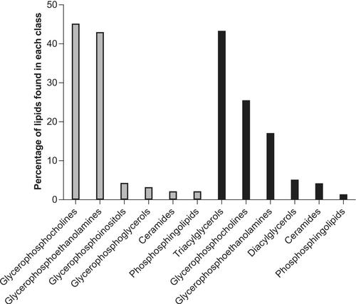
An optimised embedding method was developed for longitudinal cross-sectioning of individual worms. To obtain longitudinal cross sections, nematodes were frozen at −80°C so that all worms were rod shaped. Then, the samples were defrosted and transferred to a 10% gelatine matrix in a cryomold; the worms aligned, and the whole cryomold submerged in liquid nitrogen. Using MALDI-MSI, we were able to observe the distribution of lipids within C. elegans, with clear differences in their spatial distribution at a resolution of 5 μm. To confirm the lipids from MALDI-MSI, age-matched nematodes were subjected to LC-MS/MS. Here, 520 lipids were identified using LC-MS/MS, indicating overlap with MALDI-MSI data.

Conclusions

This optimised sample preparation technique enabled (un)targeted analysis of spatially distributed lipids within individual nematodes. The possibility to detect other biomolecules using this method thus laid the basis for prospective preclinical and toxicological studies on C. elegans.
Abstract Image

基质辅助激光解吸电离质谱成像与液相色谱-串联质谱联用技术,用于观察整条秀丽隐杆线虫体内脂质的空间分布。
原理:基质辅助激光解吸电离质谱成像(MALDI-MSI)是一种强大的无标记技术,可用于检测各种生物物种组织切片中的生物大分子(如脂质)。然而,尽管 MALDI-MSI 在许多应用中都很有用,但线虫却没有被常规地与 MALDI-MSI 结合使用。缺乏对线虫体内生物大分子空间分布的研究可能是由于样品制备方面的挑战:本研究开发了一种完整线虫的样品制备方法,评估方法是将线虫嵌入 10%明胶溶液中冷冻切片,获得纵向横截面。然后使用 RapifleX Tissuetyper 在正负极性下对切片进行 MALDI-MSI。此外,还使用与 HPLC Vanquish 系统相连的 Exploris 480 对样品进行液相色谱-串联质谱(LC-MS/MS)分析,以确认 MALDI-MSI 结果:开发了一种优化的包埋方法,用于单个蠕虫的纵向横截面。为获得纵向横截面,线虫在-80°C下冷冻,使所有蠕虫呈棒状。然后,将样本解冻并转移到低温模中的 10%明胶基质中;将蠕虫对齐,并将整个低温模浸没在液氮中。利用 MALDI-MSI,我们能够观察到虫体内脂质的分布,在 5 μm 的分辨率下,脂质的空间分布存在明显差异。为了确认 MALDI-MSI 中的脂质,我们对年龄匹配的线虫进行了 LC-MS/MS。在这里,利用 LC-MS/MS 鉴定出了 520 种脂质,表明与 MALDI-MSI 数据有重叠:结论:这种优化的样品制备技术能够对线虫个体内部空间分布的脂质进行(非)靶向分析。因此,利用这种方法检测其他生物大分子的可能性为对线虫进行前瞻性临床前研究和毒理学研究奠定了基础。
本文章由计算机程序翻译,如有差异,请以英文原文为准。
求助全文
约1分钟内获得全文 求助全文
来源期刊
CiteScore
4.10
自引率
5.00%
发文量
219
审稿时长
2.6 months
期刊介绍: Rapid Communications in Mass Spectrometry is a journal whose aim is the rapid publication of original research results and ideas on all aspects of the science of gas-phase ions; it covers all the associated scientific disciplines. There is no formal limit on paper length ("rapid" is not synonymous with "brief"), but papers should be of a length that is commensurate with the importance and complexity of the results being reported. Contributions may be theoretical or practical in nature; they may deal with methods, techniques and applications, or with the interpretation of results; they may cover any area in science that depends directly on measurements made upon gaseous ions or that is associated with such measurements.
×
引用
GB/T 7714-2015
复制
MLA
复制
APA
复制
导出至
BibTeX EndNote RefMan NoteFirst NoteExpress
×
提示
您的信息不完整,为了账户安全,请先补充。
现在去补充
×
提示
您因"违规操作"
具体请查看互助需知
我知道了
×
提示
确定
请完成安全验证×
copy
已复制链接
快去分享给好友吧!
我知道了
右上角分享
点击右上角分享
0
联系我们:info@booksci.cn Book学术提供免费学术资源搜索服务,方便国内外学者检索中英文文献。致力于提供最便捷和优质的服务体验。 Copyright © 2023 布克学术 All rights reserved.
京ICP备2023020795号-1
ghs 京公网安备 11010802042870号
Book学术文献互助
Book学术文献互助群
群 号:481959085
Book学术官方微信