Koji Nukuto, Tom Gale, Tetsuya Yamamoto, Kohei Kamada, James J. Irrgang, Volker Musahl, William Anderst
{"title":"Reliability and changes in knee cartilage T2 relaxation time from 6 to 24 months after anatomic anterior cruciate ligament reconstruction","authors":"Koji Nukuto, Tom Gale, Tetsuya Yamamoto, Kohei Kamada, James J. Irrgang, Volker Musahl, William Anderst","doi":"10.1002/jor.25939","DOIUrl":null,"url":null,"abstract":"<p>The objectives of this study were to evaluate the reliability of cartilage T2 relaxation time measurements and to identify focal changes in T2 relaxation on the affected knee from 6 to 24 months after anatomic anterior cruciate ligament reconstruction (ACLR). Data from 41 patients who received anatomic ACLR were analyzed. A bilateral 3.0-T MRI was acquired 6 and 24 months after ACLR. T2 relaxation time was measured in subregions of the femoral condyle and the tibial plateau. The root-mean-square coefficient of variation (RMS<sub>CV</sub>) was calculated to evaluate the reliability of T2 relaxation time in the contralateral knee. Subregion changes in the affected knee T2 relaxation time were identified using the contralateral knee as a reference. The superficial and full thickness layers of the central and inner regions showed good reliability. Conversely, the outer regions on the femoral side and regions in the deep layers showed poor reliability. T2 relaxation time increased in only 3 regions on the affected knee when controlling for changes in the contralateral knee, while changes in T2 relaxation time were identified in 14 regions when not using the contralateral knee as a reference. In conclusion, evaluation of cartilage degeneration by T2 relaxation time after ACLR is most reliable for central and inner cartilage regions. Cartilage degeneration occurs in the central and outer regions of the lateral femoral condyle from 6 to 24 months after anatomic ACLR.</p>","PeriodicalId":16650,"journal":{"name":"Journal of Orthopaedic Research®","volume":"42 12","pages":"2683-2692"},"PeriodicalIF":2.1000,"publicationDate":"2024-07-20","publicationTypes":"Journal Article","fieldsOfStudy":null,"isOpenAccess":false,"openAccessPdf":"https://onlinelibrary.wiley.com/doi/epdf/10.1002/jor.25939","citationCount":"0","resultStr":null,"platform":"Semanticscholar","paperid":null,"PeriodicalName":"Journal of Orthopaedic Research®","FirstCategoryId":"3","ListUrlMain":"https://onlinelibrary.wiley.com/doi/10.1002/jor.25939","RegionNum":3,"RegionCategory":"医学","ArticlePicture":[],"TitleCN":null,"AbstractTextCN":null,"PMCID":null,"EPubDate":"","PubModel":"","JCR":"Q2","JCRName":"ORTHOPEDICS","Score":null,"Total":0}
引用次数: 0
Abstract
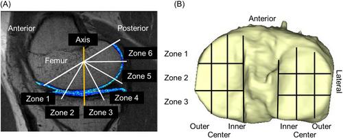
The objectives of this study were to evaluate the reliability of cartilage T2 relaxation time measurements and to identify focal changes in T2 relaxation on the affected knee from 6 to 24 months after anatomic anterior cruciate ligament reconstruction (ACLR). Data from 41 patients who received anatomic ACLR were analyzed. A bilateral 3.0-T MRI was acquired 6 and 24 months after ACLR. T2 relaxation time was measured in subregions of the femoral condyle and the tibial plateau. The root-mean-square coefficient of variation (RMSCV) was calculated to evaluate the reliability of T2 relaxation time in the contralateral knee. Subregion changes in the affected knee T2 relaxation time were identified using the contralateral knee as a reference. The superficial and full thickness layers of the central and inner regions showed good reliability. Conversely, the outer regions on the femoral side and regions in the deep layers showed poor reliability. T2 relaxation time increased in only 3 regions on the affected knee when controlling for changes in the contralateral knee, while changes in T2 relaxation time were identified in 14 regions when not using the contralateral knee as a reference. In conclusion, evaluation of cartilage degeneration by T2 relaxation time after ACLR is most reliable for central and inner cartilage regions. Cartilage degeneration occurs in the central and outer regions of the lateral femoral condyle from 6 to 24 months after anatomic ACLR.
期刊介绍:
The Journal of Orthopaedic Research is the forum for the rapid publication of high quality reports of new information on the full spectrum of orthopaedic research, including life sciences, engineering, translational, and clinical studies.

 求助内容:
求助内容: 应助结果提醒方式:


