Astragalus mongholicus bunge and panax notoginseng formula (A&P) improves renal fibrosis in UUO mice via inhibiting the long non-coding RNA A330074K22Rik and downregulating ferroptosis signaling.
IF 3.3 2区 医学Q1 INTEGRATIVE & COMPLEMENTARY MEDICINE
Xia Zhong, Yue Huang, Jian Jia, Jian Liu, Hongwei Su, Qiongdan Hu, Ruizhi Tan, Li Wang
{"title":"Astragalus mongholicus bunge and panax notoginseng formula (A&P) improves renal fibrosis in UUO mice via inhibiting the long non-coding RNA A330074K22Rik and downregulating ferroptosis signaling.","authors":"Xia Zhong, Yue Huang, Jian Jia, Jian Liu, Hongwei Su, Qiongdan Hu, Ruizhi Tan, Li Wang","doi":"10.1186/s12906-024-04557-4","DOIUrl":null,"url":null,"abstract":"<p><strong>Background: </strong>Chronic kidney disease (CKD) and its associated end-stage renal disease (ESRD) are significant health problems that pose a threat to human well-being. Renal fibrosis is a common feature and ultimate pathological outcome of various CKD leading to ESRD. The Astragalus mongholicus Bunge and Panax notoginseng formula (A&P) is a refined compound formulated by our research group, which has been clinically administered for over a decade and has demonstrated the ability to improve the inflammatory state of various acute or chronic kidney diseases. However, the underlying mechanism by which A&P ameliorates renal fibrosis remains unclear.</p><p><strong>Methods: </strong>We established a mouse model by surgically ligating the unilateral ureter to induce renal injury in vivo. And we utilized renal in situ electroporation of a plasmid with low LncRNA A33 expression to establish the unilateral ureteral obstruction(UUO)mouse model. In vitro, we stimulated primary tubular epithelial cells(pTEC) injury using TGF-β1, siRNA-A33, and pcDNA3.1-A33 plasmids were transfected into pTECs to respectively knockdown and overexpress LncRNA A33, and both in vitro and in vivo models were intervened with A&P.</p><p><strong>Results: </strong>The results demonstrated that A&P effectively alleviated renal fibrosis in mice. Subsequent findings indicated high expression of LncRNA A33 in the kidneys of UUO mice and TGF-β1-induced renal tubular cells. In situ, renal electroporation of a plasmid with reduced LncRNA A33 expression revealed that inhibiting LncRNA A33 significantly improved renal fibrosis in UUO mice. Moreover, A&P effectively suppressed LncRNA A33 expression both in vitro and in vivo. Subsequent downregulation of LncRNA A33 in renal tubular epithelial cells resulted in the downregulation of numerous fibrotic markers, a significant inhibition of LncRNA A33, and a notable reduction in downstream ferroptosis signaling. Cell experiments demonstrated that A&P improved renal fibrosis in UUO mice by inhibiting LncRNA A33 and downregulating ferroptosis signaling.</p><p><strong>Conclusion: </strong>Through the inhibition of LncRNA A33 and subsequent downregulation of ferroptosis signaling, A&P showed potential as a therapeutic approach for improving renal fibrosis in UUO mice, providing a potential treatment avenue for CKD.</p>","PeriodicalId":9128,"journal":{"name":"BMC Complementary Medicine and Therapies","volume":null,"pages":null},"PeriodicalIF":3.3000,"publicationDate":"2024-07-19","publicationTypes":"Journal Article","fieldsOfStudy":null,"isOpenAccess":false,"openAccessPdf":"https://www.ncbi.nlm.nih.gov/pmc/articles/PMC11264518/pdf/","citationCount":"0","resultStr":null,"platform":"Semanticscholar","paperid":null,"PeriodicalName":"BMC Complementary Medicine and Therapies","FirstCategoryId":"3","ListUrlMain":"https://doi.org/10.1186/s12906-024-04557-4","RegionNum":2,"RegionCategory":"医学","ArticlePicture":[],"TitleCN":null,"AbstractTextCN":null,"PMCID":null,"EPubDate":"","PubModel":"","JCR":"Q1","JCRName":"INTEGRATIVE & COMPLEMENTARY MEDICINE","Score":null,"Total":0}
引用次数: 0
Abstract
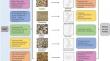
Background: Chronic kidney disease (CKD) and its associated end-stage renal disease (ESRD) are significant health problems that pose a threat to human well-being. Renal fibrosis is a common feature and ultimate pathological outcome of various CKD leading to ESRD. The Astragalus mongholicus Bunge and Panax notoginseng formula (A&P) is a refined compound formulated by our research group, which has been clinically administered for over a decade and has demonstrated the ability to improve the inflammatory state of various acute or chronic kidney diseases. However, the underlying mechanism by which A&P ameliorates renal fibrosis remains unclear.
Methods: We established a mouse model by surgically ligating the unilateral ureter to induce renal injury in vivo. And we utilized renal in situ electroporation of a plasmid with low LncRNA A33 expression to establish the unilateral ureteral obstruction(UUO)mouse model. In vitro, we stimulated primary tubular epithelial cells(pTEC) injury using TGF-β1, siRNA-A33, and pcDNA3.1-A33 plasmids were transfected into pTECs to respectively knockdown and overexpress LncRNA A33, and both in vitro and in vivo models were intervened with A&P.
Results: The results demonstrated that A&P effectively alleviated renal fibrosis in mice. Subsequent findings indicated high expression of LncRNA A33 in the kidneys of UUO mice and TGF-β1-induced renal tubular cells. In situ, renal electroporation of a plasmid with reduced LncRNA A33 expression revealed that inhibiting LncRNA A33 significantly improved renal fibrosis in UUO mice. Moreover, A&P effectively suppressed LncRNA A33 expression both in vitro and in vivo. Subsequent downregulation of LncRNA A33 in renal tubular epithelial cells resulted in the downregulation of numerous fibrotic markers, a significant inhibition of LncRNA A33, and a notable reduction in downstream ferroptosis signaling. Cell experiments demonstrated that A&P improved renal fibrosis in UUO mice by inhibiting LncRNA A33 and downregulating ferroptosis signaling.
Conclusion: Through the inhibition of LncRNA A33 and subsequent downregulation of ferroptosis signaling, A&P showed potential as a therapeutic approach for improving renal fibrosis in UUO mice, providing a potential treatment avenue for CKD.
 求助内容:
求助内容: 应助结果提醒方式:


