{"title":"Free iron accumulation and oxidative stress burden induce ferroptotic atrophy of chicken yolk sac during the late embryogenesis","authors":"Huichao Liu, Zehe Song, Xi He, Haihan Zhang","doi":"10.1002/aro2.74","DOIUrl":null,"url":null,"abstract":"<p>The aim of this study was to investigate the mechanism of iron homeostasis and the ferroptosis pathway for yolk sac atrophy during late embryogenesis. To study the mechanism of yolk sac atrophy, 100 eggs were used. Further, 500 eggs were randomly divided into five treatments and in ovo feeding with different iron sources, such as FeSO<sub>4</sub>, ferrous glycinate (Fe-Gly), or deferoxamine (DFO), to study the effects of free iron content on hatching quality and embryonic development. The results showed that total iron content of yolk decreased, but yolk sac increased from embryonic(E)13 to E19 (<i>p</i> < 0.05). Comparison of gene expression of iron transport systems showed that free iron accumulation and dysfunction occurred in the yolk sac. Yolk sac metabolites at E19 compared to E13 were more enriched in histidine and sulfur pathways, suppressing glutathione synthesis and resulting in oxidative stress damage in the yolk sac. Combined analysis of differential metabolites and gene expression in ferroptosis pathway at E13 and E19 revealed the activation of the yolk sac during late embryogenesis was probably through up-regulation of <i>ACSL4</i> expression and down-regulation of <i>GPX4</i> expression. Furthermore, in ovo feeding FeSO<sub>4</sub> shortened the incubation time compared to CON, while Fe-Gly or DFO delayed the hatching peak and increased hatching weight with less residual yolk. Collectively, it can be concluded that yolk sac atrophy during late embryogenesis may be mediated by iron disorders and provides a novel insight to modulate yolk sac nutrition, and hatching efficiency in chickens.</p>","PeriodicalId":100086,"journal":{"name":"Animal Research and One Health","volume":"2 3","pages":"285-299"},"PeriodicalIF":0.0000,"publicationDate":"2024-07-08","publicationTypes":"Journal Article","fieldsOfStudy":null,"isOpenAccess":false,"openAccessPdf":"https://onlinelibrary.wiley.com/doi/epdf/10.1002/aro2.74","citationCount":"0","resultStr":null,"platform":"Semanticscholar","paperid":null,"PeriodicalName":"Animal Research and One Health","FirstCategoryId":"1085","ListUrlMain":"https://onlinelibrary.wiley.com/doi/10.1002/aro2.74","RegionNum":0,"RegionCategory":null,"ArticlePicture":[],"TitleCN":null,"AbstractTextCN":null,"PMCID":null,"EPubDate":"","PubModel":"","JCR":"","JCRName":"","Score":null,"Total":0}
引用次数: 0
Abstract
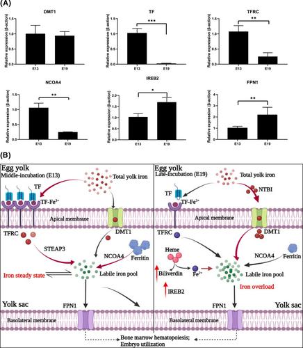
The aim of this study was to investigate the mechanism of iron homeostasis and the ferroptosis pathway for yolk sac atrophy during late embryogenesis. To study the mechanism of yolk sac atrophy, 100 eggs were used. Further, 500 eggs were randomly divided into five treatments and in ovo feeding with different iron sources, such as FeSO4, ferrous glycinate (Fe-Gly), or deferoxamine (DFO), to study the effects of free iron content on hatching quality and embryonic development. The results showed that total iron content of yolk decreased, but yolk sac increased from embryonic(E)13 to E19 (p < 0.05). Comparison of gene expression of iron transport systems showed that free iron accumulation and dysfunction occurred in the yolk sac. Yolk sac metabolites at E19 compared to E13 were more enriched in histidine and sulfur pathways, suppressing glutathione synthesis and resulting in oxidative stress damage in the yolk sac. Combined analysis of differential metabolites and gene expression in ferroptosis pathway at E13 and E19 revealed the activation of the yolk sac during late embryogenesis was probably through up-regulation of ACSL4 expression and down-regulation of GPX4 expression. Furthermore, in ovo feeding FeSO4 shortened the incubation time compared to CON, while Fe-Gly or DFO delayed the hatching peak and increased hatching weight with less residual yolk. Collectively, it can be concluded that yolk sac atrophy during late embryogenesis may be mediated by iron disorders and provides a novel insight to modulate yolk sac nutrition, and hatching efficiency in chickens.

 求助内容:
求助内容: 应助结果提醒方式:


