Advancements of non‐invasive imaging technologies for the diagnosis and staging of liver fibrosis: Present and future
引用次数: 0
Abstract
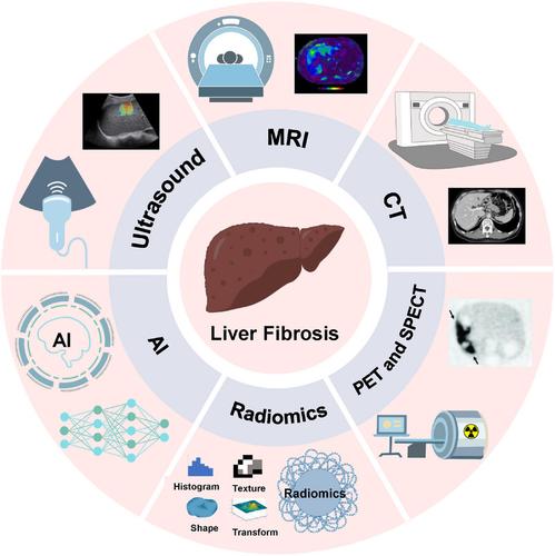
Liver fibrosis is a reparative response triggered by liver injury. Non‐invasive assessment and staging of liver fibrosis in patients with chronic liver disease are of paramount importance, as treatment strategies and prognoses depend significantly on the degree of fibrosis. Although liver fibrosis has traditionally been staged through invasive liver biopsy, this method is prone to sampling errors, particularly when biopsy sizes are inadequate. Consequently, there is an urgent clinical need for an alternative to biopsy, one that ensures precise, sensitive, and non‐invasive diagnosis and staging of liver fibrosis. Non‐invasive imaging assessments have assumed a pivotal role in clinical practice, enjoying growing popularity and acceptance due to their potential for diagnosing, staging, and monitoring liver fibrosis. In this comprehensive review, we first delved into the current landscape of non‐invasive imaging technologies, assessing their accuracy and the transformative impact they have had on the diagnosis and management of liver fibrosis in both clinical practice and animal models. Additionally, we provided an in‐depth exploration of recent advancements in ultrasound imaging, computed tomography imaging, magnetic resonance imaging, nuclear medicine imaging, radiomics, and artificial intelligence within the field of liver fibrosis research. We summarized the key concepts, advantages, limitations, and diagnostic performance of each technique. Finally, we discussed the challenges associated with clinical implementation and offer our perspective on advancing the field, hoping to provide alternative directions for the future research.
用于肝纤维化诊断和分期的无创成像技术的进展:现状与未来
肝纤维化是肝损伤引发的一种修复反应。对慢性肝病患者的肝纤维化进行无创评估和分期至关重要,因为治疗策略和预后在很大程度上取决于肝纤维化的程度。虽然肝纤维化传统上是通过侵入性肝活检来分期的,但这种方法容易出现取样误差,尤其是活检样本量不足时。因此,临床上迫切需要一种可替代活检的方法,以确保对肝纤维化进行精确、灵敏和无创的诊断和分期。无创成像评估在临床实践中发挥着举足轻重的作用,因其在诊断、分期和监测肝纤维化方面的潜力而日益受到欢迎和认可。在这篇综合性综述中,我们首先深入探讨了无创成像技术的现状,评估了其准确性以及在临床实践和动物模型中对肝纤维化诊断和管理产生的变革性影响。此外,我们还深入探讨了肝纤维化研究领域中超声成像、计算机断层扫描成像、磁共振成像、核医学成像、放射组学和人工智能的最新进展。我们总结了每种技术的关键概念、优势、局限性和诊断性能。最后,我们讨论了与临床实施相关的挑战,并提出了我们对推进该领域发展的看法,希望能为未来的研究提供可供选择的方向。
本文章由计算机程序翻译,如有差异,请以英文原文为准。
求助全文
约1分钟内获得全文
求助全文

 求助内容:
求助内容: 应助结果提醒方式:


