Caterina Redwanz , Ricardo H. Pires , Doreen Biedenweg , Stefan Groß , Oliver Otto , Stephanie Könemann
{"title":"Endothelin-1 influences mechanical properties and contractility of hiPSC derived cardiomyocytes resulting in diastolic dysfunction","authors":"Caterina Redwanz , Ricardo H. Pires , Doreen Biedenweg , Stefan Groß , Oliver Otto , Stephanie Könemann","doi":"10.1016/j.yjmcc.2024.07.004","DOIUrl":null,"url":null,"abstract":"<div><p>A better understanding of the underlying pathomechanisms of diastolic dysfunction is crucial for the development of targeted therapeutic options with the aim to increase the patients' quality of life. In order to shed light on the processes involved, suitable models are required. Here, effects of endothelin-1 (ET-1) treatment on cardiomyocytes derived from human induced pluripotent stem cells (hiPSCs) were investigated. While it is well established, that ET-1 treatment induces hypertrophy in cardiomyocytes, resulting changes in cell mechanics and contractile behavior with focus on relaxation have not been examined before. Cardiomyocytes were treated with 10 nM of ET-1 for 24 h and 48 h, respectively. Hypertrophy was confirmed by real-time deformability cytometry (RT-DC) which was also used to assess the mechanical properties of cardiomyocytes. For investigation of the contractile behavior, 24 h phase contrast video microscopy was applied. To get a deeper insight into changes on the molecular biological level, gene expression analysis was performed using the NanoString nCounter® cardiovascular disease panel. Besides an increased cell size, ET-1 treated cardiomyocytes are stiffer and show an impaired relaxation. Gene expression patterns in ET-1 treated hiPSC derived cardiomyocytes showed that pathways associated with cardiovascular diseases, cardiac hypertrophy and extracellular matrix were upregulated while those associated with fatty acid metabolism were downregulated. We conclude that alterations in cardiomyocytes after ET-1 treatment go far beyond hypertrophy and represent a useful model for diastolic dysfunction.</p></div>","PeriodicalId":16402,"journal":{"name":"Journal of molecular and cellular cardiology","volume":"194 ","pages":"Pages 105-117"},"PeriodicalIF":4.9000,"publicationDate":"2024-07-15","publicationTypes":"Journal Article","fieldsOfStudy":null,"isOpenAccess":false,"openAccessPdf":"https://www.sciencedirect.com/science/article/pii/S0022282824001184/pdfft?md5=a49d2c318127024240184c8ba386620a&pid=1-s2.0-S0022282824001184-main.pdf","citationCount":"0","resultStr":null,"platform":"Semanticscholar","paperid":null,"PeriodicalName":"Journal of molecular and cellular cardiology","FirstCategoryId":"3","ListUrlMain":"https://www.sciencedirect.com/science/article/pii/S0022282824001184","RegionNum":2,"RegionCategory":"医学","ArticlePicture":[],"TitleCN":null,"AbstractTextCN":null,"PMCID":null,"EPubDate":"","PubModel":"","JCR":"Q1","JCRName":"CARDIAC & CARDIOVASCULAR SYSTEMS","Score":null,"Total":0}
引用次数: 0
Abstract
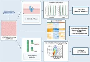
A better understanding of the underlying pathomechanisms of diastolic dysfunction is crucial for the development of targeted therapeutic options with the aim to increase the patients' quality of life. In order to shed light on the processes involved, suitable models are required. Here, effects of endothelin-1 (ET-1) treatment on cardiomyocytes derived from human induced pluripotent stem cells (hiPSCs) were investigated. While it is well established, that ET-1 treatment induces hypertrophy in cardiomyocytes, resulting changes in cell mechanics and contractile behavior with focus on relaxation have not been examined before. Cardiomyocytes were treated with 10 nM of ET-1 for 24 h and 48 h, respectively. Hypertrophy was confirmed by real-time deformability cytometry (RT-DC) which was also used to assess the mechanical properties of cardiomyocytes. For investigation of the contractile behavior, 24 h phase contrast video microscopy was applied. To get a deeper insight into changes on the molecular biological level, gene expression analysis was performed using the NanoString nCounter® cardiovascular disease panel. Besides an increased cell size, ET-1 treated cardiomyocytes are stiffer and show an impaired relaxation. Gene expression patterns in ET-1 treated hiPSC derived cardiomyocytes showed that pathways associated with cardiovascular diseases, cardiac hypertrophy and extracellular matrix were upregulated while those associated with fatty acid metabolism were downregulated. We conclude that alterations in cardiomyocytes after ET-1 treatment go far beyond hypertrophy and represent a useful model for diastolic dysfunction.
期刊介绍:
The Journal of Molecular and Cellular Cardiology publishes work advancing knowledge of the mechanisms responsible for both normal and diseased cardiovascular function. To this end papers are published in all relevant areas. These include (but are not limited to): structural biology; genetics; proteomics; morphology; stem cells; molecular biology; metabolism; biophysics; bioengineering; computational modeling and systems analysis; electrophysiology; pharmacology and physiology. Papers are encouraged with both basic and translational approaches. The journal is directed not only to basic scientists but also to clinical cardiologists who wish to follow the rapidly advancing frontiers of basic knowledge of the heart and circulation.
 求助内容:
求助内容: 应助结果提醒方式:


