Wenying Wang, Li Peng, Lian He, Yan Chen, Mingshan Jiang, Xue Luo, Guoqiang Gao
{"title":"Applicability of combined high-frequency and contrast-enhanced ultrasound in finger extensor tendon injuries: three case reports.","authors":"Wenying Wang, Li Peng, Lian He, Yan Chen, Mingshan Jiang, Xue Luo, Guoqiang Gao","doi":"10.1186/s13089-024-00376-3","DOIUrl":null,"url":null,"abstract":"<p><strong>Background: </strong>By combining high-frequency and contrast-enhanced ultrasound (CEUS), the position of the severed end of a finger extensor tendon injury and the injury classification can be determined as part of a comprehensive preoperative evaluation in clinical practice. However, there have been no reports of high-frequency ultrasound combined with CEUS for the preoperative diagnosis of human finger extensor tendon injury.</p><p><strong>Cases presentation: </strong>One case of complete rupture of the extensor tendon was diagnosed by ultrasound, which was completely consistent with the surgery; one case of incomplete rupture was ultimately confirmed clinically; and one case of distal phalangeal bone base avulsion fracture with tendon contusion and missed diagnosis on the first radiographic examination was confirmed by follow-up radiographic examination.</p><p><strong>Conclusions: </strong>Different types of finger extensor tendon injuries exhibit distinctive contrast-enhanced ultrasonography findings. Combined high-frequency and contrast-enhanced ultrasound can accurately locate the position of the severed end of the finger extensor tendon injury before surgery while observing the contrast agent filling area to clarify injury classification, providing a reliable imaging basis for clinical practice and ultimately developing personalized diagnosis and treatment plans for patients to ensure minimal trauma and pain, as well as optimal treatment effects.</p>","PeriodicalId":36911,"journal":{"name":"Ultrasound Journal","volume":"16 1","pages":"36"},"PeriodicalIF":2.9000,"publicationDate":"2024-07-17","publicationTypes":"Journal Article","fieldsOfStudy":null,"isOpenAccess":false,"openAccessPdf":"https://www.ncbi.nlm.nih.gov/pmc/articles/PMC11254872/pdf/","citationCount":"0","resultStr":null,"platform":"Semanticscholar","paperid":null,"PeriodicalName":"Ultrasound Journal","FirstCategoryId":"1085","ListUrlMain":"https://doi.org/10.1186/s13089-024-00376-3","RegionNum":0,"RegionCategory":null,"ArticlePicture":[],"TitleCN":null,"AbstractTextCN":null,"PMCID":null,"EPubDate":"","PubModel":"","JCR":"Q2","JCRName":"Medicine","Score":null,"Total":0}
引用次数: 0
Abstract
Background: By combining high-frequency and contrast-enhanced ultrasound (CEUS), the position of the severed end of a finger extensor tendon injury and the injury classification can be determined as part of a comprehensive preoperative evaluation in clinical practice. However, there have been no reports of high-frequency ultrasound combined with CEUS for the preoperative diagnosis of human finger extensor tendon injury.
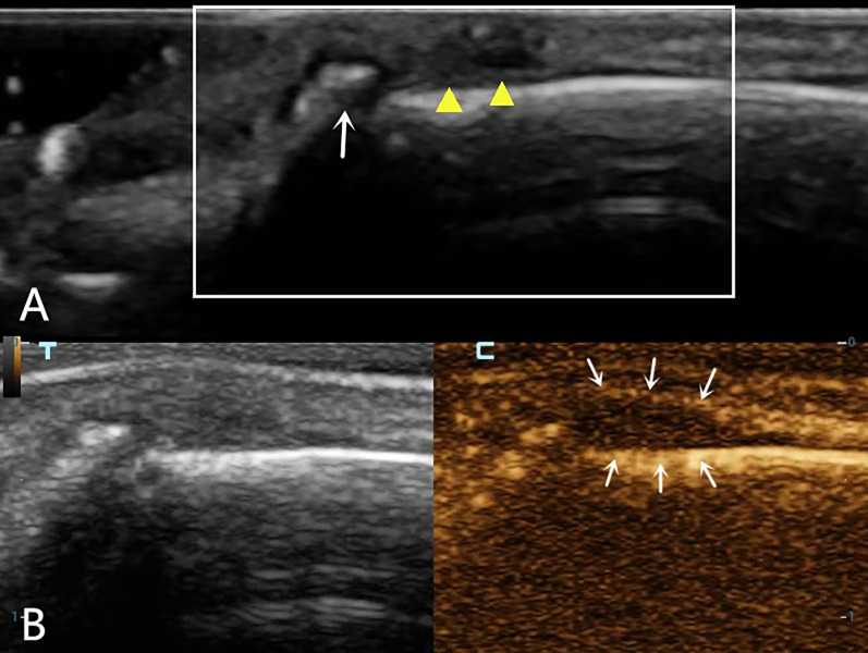
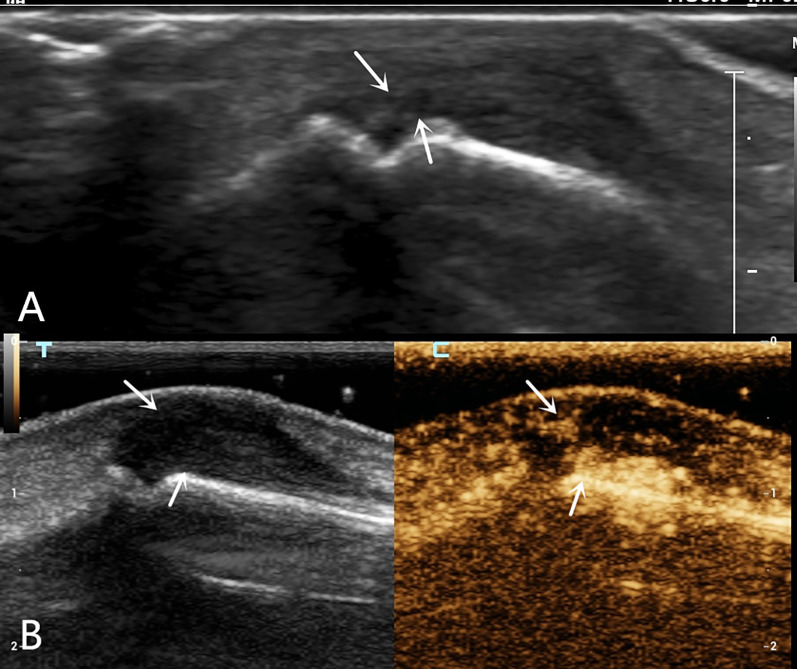
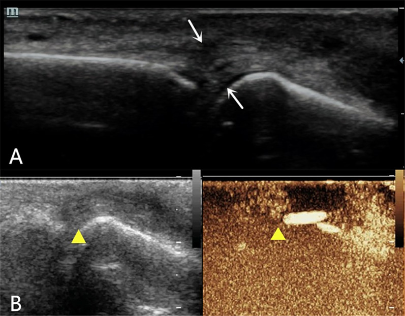
Cases presentation: One case of complete rupture of the extensor tendon was diagnosed by ultrasound, which was completely consistent with the surgery; one case of incomplete rupture was ultimately confirmed clinically; and one case of distal phalangeal bone base avulsion fracture with tendon contusion and missed diagnosis on the first radiographic examination was confirmed by follow-up radiographic examination.
Conclusions: Different types of finger extensor tendon injuries exhibit distinctive contrast-enhanced ultrasonography findings. Combined high-frequency and contrast-enhanced ultrasound can accurately locate the position of the severed end of the finger extensor tendon injury before surgery while observing the contrast agent filling area to clarify injury classification, providing a reliable imaging basis for clinical practice and ultimately developing personalized diagnosis and treatment plans for patients to ensure minimal trauma and pain, as well as optimal treatment effects.

 求助内容:
求助内容: 应助结果提醒方式:


