{"title":"Association of tibial slope alterations with anterior cruciate ligament (ACL) injury and mucoid degeneration.","authors":"Varun Ravi, Mahad Rehman, Shuda Xia, Avneesh Chhabra, Flavio Duarte Silva","doi":"10.1007/s00256-024-04744-x","DOIUrl":null,"url":null,"abstract":"<p><strong>Objective: </strong>To compare radiographic measurements of lateral tibial slope (LTS), medial TS (MTS), and coronal TS (CTS) in MRI-defined intact, injured, and mucoid-degenerated native ACL knees and determine inter-reader reliability.</p><p><strong>Materials and methods: </strong>Patient records from 2 years at tertiary care hospitals were reviewed for individuals aged 18-100 undergoing 3-Tesla knee MRI and radiographs. Two randomly selected cohorts, control, and pathologic ACLs on MRI with 86 patients each, were age, gender, and BMI-matched. A fellowship-trained musculoskeletal radiologist reevaluated curated images, characterizing ACL status. Two trained medical students independently collected clinical data and measured slopes on blinded radiographs. ICC, Cohen's kappa, and case-control matching were performed using SPSS statistical package, with ICC and ANOVA used for comparisons.</p><p><strong>Results: </strong>Among 172 patients with 172 MRIs and radiographs, there were 86 controls and 86 ACL lesions. There were 108/172 (62.79%) males and 64/172 (37.21%) females. ICCs were 0.966 for MTS, 0.975 for LTS, and 0.978 for CTS. Mucoid degeneration patients had a higher BMI and were older than control (p < .05) or completely torn (p < .001) ACL patients. There was no difference in TS between normal and pathologic ACLs; however, LTS-MTS differences were larger with partial tears (2.5 ± 4.9) than normal ACLs by 4.5° (± 1.2, p < .001), complete tears by 4.5° (± 1.3, p < .001), and mucoid degeneration by 4.9° (± 1.5, p = .001).</p><p><strong>Conclusion: </strong>Various TS measurements are reliable. LTS-MTS differences are associated with different ACL lesions compared to normal ACLs.</p>","PeriodicalId":21783,"journal":{"name":"Skeletal Radiology","volume":" ","pages":"325-334"},"PeriodicalIF":1.9000,"publicationDate":"2025-02-01","publicationTypes":"Journal Article","fieldsOfStudy":null,"isOpenAccess":false,"openAccessPdf":"","citationCount":"0","resultStr":null,"platform":"Semanticscholar","paperid":null,"PeriodicalName":"Skeletal Radiology","FirstCategoryId":"3","ListUrlMain":"https://doi.org/10.1007/s00256-024-04744-x","RegionNum":3,"RegionCategory":"医学","ArticlePicture":[],"TitleCN":null,"AbstractTextCN":null,"PMCID":null,"EPubDate":"2024/7/17 0:00:00","PubModel":"Epub","JCR":"Q2","JCRName":"ORTHOPEDICS","Score":null,"Total":0}
引用次数: 0
Abstract
Objective: To compare radiographic measurements of lateral tibial slope (LTS), medial TS (MTS), and coronal TS (CTS) in MRI-defined intact, injured, and mucoid-degenerated native ACL knees and determine inter-reader reliability.
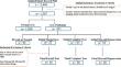
Materials and methods: Patient records from 2 years at tertiary care hospitals were reviewed for individuals aged 18-100 undergoing 3-Tesla knee MRI and radiographs. Two randomly selected cohorts, control, and pathologic ACLs on MRI with 86 patients each, were age, gender, and BMI-matched. A fellowship-trained musculoskeletal radiologist reevaluated curated images, characterizing ACL status. Two trained medical students independently collected clinical data and measured slopes on blinded radiographs. ICC, Cohen's kappa, and case-control matching were performed using SPSS statistical package, with ICC and ANOVA used for comparisons.
Results: Among 172 patients with 172 MRIs and radiographs, there were 86 controls and 86 ACL lesions. There were 108/172 (62.79%) males and 64/172 (37.21%) females. ICCs were 0.966 for MTS, 0.975 for LTS, and 0.978 for CTS. Mucoid degeneration patients had a higher BMI and were older than control (p < .05) or completely torn (p < .001) ACL patients. There was no difference in TS between normal and pathologic ACLs; however, LTS-MTS differences were larger with partial tears (2.5 ± 4.9) than normal ACLs by 4.5° (± 1.2, p < .001), complete tears by 4.5° (± 1.3, p < .001), and mucoid degeneration by 4.9° (± 1.5, p = .001).
Conclusion: Various TS measurements are reliable. LTS-MTS differences are associated with different ACL lesions compared to normal ACLs.
期刊介绍:
Skeletal Radiology provides a forum for the dissemination of current knowledge and information dealing with disorders of the musculoskeletal system including the spine. While emphasizing the radiological aspects of the many varied skeletal abnormalities, the journal also adopts an interdisciplinary approach, reflecting the membership of the International Skeletal Society. Thus, the anatomical, pathological, physiological, clinical, metabolic and epidemiological aspects of the many entities affecting the skeleton receive appropriate consideration.
This is the Journal of the International Skeletal Society and the Official Journal of the Society of Skeletal Radiology and the Australasian Musculoskelelal Imaging Group.

 求助内容:
求助内容: 应助结果提醒方式:


