Jordan Guntin, Ashley Ricciardelli, Alex Flores, Jeffrey Chen, Jeffrey Treiber, Alfonso Fuentes
{"title":"Pure epidural extraosseous cavernous hemangioma with thoracic myelopathy: case report and review of literature.","authors":"Jordan Guntin, Ashley Ricciardelli, Alex Flores, Jeffrey Chen, Jeffrey Treiber, Alfonso Fuentes","doi":"10.1038/s41394-024-00655-0","DOIUrl":null,"url":null,"abstract":"<p><strong>Introduction: </strong>Pure epidural spinal cavernous hemangiomas are rare, benign vascular tumors that account for approximately 4% of all spinal epidural tumors. Due to their dumbbell shape and propensity for foraminal invasion, they are often misdiagnosed and inadequately treated. We present a case of a 58-year-old male with extra-osseous cavernous hemangioma to better aid in diagnosis and management of these lesions.</p><p><strong>Case presentation: </strong>A 58-year-old male presented with chronic lower back pain, progressive lower extremity weakness, T10 sensory level, absent lower extremity proprioception, hyperreflexia, and an episode of bowel incontinence. Imaging demonstrated T7-T10 homogenous dorsal epidural mass causing cord signal change. He underwent resection with histopathologic exam revealing a pure epidural cavernous hemangioma.</p><p><strong>Conclusion: </strong>Spinal epidural cavernous hemangiomas are exceedingly rare lesions that are often misdiagnosed as nerve sheath tumors and meningiomas. Common features include chronic pain and myelopathy as well as T1 isodensity, T2 hyperintensity, and homogenous enhancement. Uniquely, they present as a lobulated, spindled shape with tapered ends in the dorsal epidural space. Both gross and subtotal resection result in favorable neurologic outcomes.</p>","PeriodicalId":22079,"journal":{"name":"Spinal Cord Series and Cases","volume":"10 1","pages":"48"},"PeriodicalIF":0.9000,"publicationDate":"2024-07-15","publicationTypes":"Journal Article","fieldsOfStudy":null,"isOpenAccess":false,"openAccessPdf":"https://www.ncbi.nlm.nih.gov/pmc/articles/PMC11251031/pdf/","citationCount":"0","resultStr":null,"platform":"Semanticscholar","paperid":null,"PeriodicalName":"Spinal Cord Series and Cases","FirstCategoryId":"1085","ListUrlMain":"https://doi.org/10.1038/s41394-024-00655-0","RegionNum":0,"RegionCategory":null,"ArticlePicture":[],"TitleCN":null,"AbstractTextCN":null,"PMCID":null,"EPubDate":"","PubModel":"","JCR":"Q4","JCRName":"CLINICAL NEUROLOGY","Score":null,"Total":0}
引用次数: 0
Abstract
Introduction: Pure epidural spinal cavernous hemangiomas are rare, benign vascular tumors that account for approximately 4% of all spinal epidural tumors. Due to their dumbbell shape and propensity for foraminal invasion, they are often misdiagnosed and inadequately treated. We present a case of a 58-year-old male with extra-osseous cavernous hemangioma to better aid in diagnosis and management of these lesions.
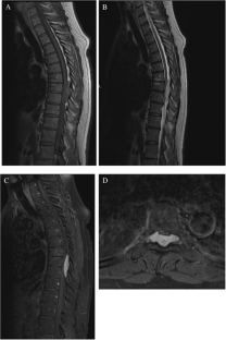
Case presentation: A 58-year-old male presented with chronic lower back pain, progressive lower extremity weakness, T10 sensory level, absent lower extremity proprioception, hyperreflexia, and an episode of bowel incontinence. Imaging demonstrated T7-T10 homogenous dorsal epidural mass causing cord signal change. He underwent resection with histopathologic exam revealing a pure epidural cavernous hemangioma.
Conclusion: Spinal epidural cavernous hemangiomas are exceedingly rare lesions that are often misdiagnosed as nerve sheath tumors and meningiomas. Common features include chronic pain and myelopathy as well as T1 isodensity, T2 hyperintensity, and homogenous enhancement. Uniquely, they present as a lobulated, spindled shape with tapered ends in the dorsal epidural space. Both gross and subtotal resection result in favorable neurologic outcomes.

 求助内容:
求助内容: 应助结果提醒方式:


