{"title":"Impact of acid and laser etching of enamel on microleakage in different adhesive systems.","authors":"Sevim Atilan Yavuz, Ayse Tugba Erturk Avunduk, Ozcan Karatas, Nazire Nurdan Çakır Kılınç, Ebru Delikan","doi":"10.1007/s10103-024-04120-0","DOIUrl":null,"url":null,"abstract":"<p><p>This study aimed to evaluate the microleakage of light-cured and self-cured adhesives on enamel surfaces selectively etched with Er, Cr: YSGG laser or 35% phosphoric acid. A total of 60 class V cavities were prepared 1 mm above the cemento-enamel junction (CEJ). The specimens were randomly divided into six groups. Group 1: Clearfil SE Bond with no conditioning, Group 2: Tokuyama Universal Bond with no conditioning, Group 3: Clearfil SE Bond conditioned with 35% phosphoric acid, Group 4: Tokuyama Universal Bond conditioned with 35% phosphoric acid, Group 5: Clearfil SE Bond conditioned with Er, Cr: YSGG laser and Group 6: Tokuyama Universal Bond conditioned with Er, Cr: YSGG laser. Microleakage was evaluated qualitatively (visually) and quantitatively (ImageJ). The data were analyzed using IBM SPSS V23 and submitted to Kruskal-Wallis and Wilcoxon tests. The significance level was set at p < 0.05. In all evaluation methods, the microleakage scores exhibit significant differences (p*<0.001). Group 1 and Group 3 exhibited similar and lower microleakage values than the Group 5. In the occlusal margin, the microleakage values were similar in Group 2, Group 4, and Group 6, whereas in the gingival margin Group 4 showed significantly lower leakage compared to Group 2. Regardless of the etching protocols and adhesive systems used, less microleakage was observed on the occlusal surface than on the gingival surface. Phosphoric acid etching provides better results than laser etching for enamel surface treatment on both occlusal and gingival surfaces.</p>","PeriodicalId":17978,"journal":{"name":"Lasers in Medical Science","volume":null,"pages":null},"PeriodicalIF":2.1000,"publicationDate":"2024-07-15","publicationTypes":"Journal Article","fieldsOfStudy":null,"isOpenAccess":false,"openAccessPdf":"https://www.ncbi.nlm.nih.gov/pmc/articles/PMC11249556/pdf/","citationCount":"0","resultStr":null,"platform":"Semanticscholar","paperid":null,"PeriodicalName":"Lasers in Medical Science","FirstCategoryId":"5","ListUrlMain":"https://doi.org/10.1007/s10103-024-04120-0","RegionNum":4,"RegionCategory":"医学","ArticlePicture":[],"TitleCN":null,"AbstractTextCN":null,"PMCID":null,"EPubDate":"","PubModel":"","JCR":"Q3","JCRName":"ENGINEERING, BIOMEDICAL","Score":null,"Total":0}
引用次数: 0
Abstract
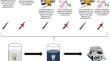
This study aimed to evaluate the microleakage of light-cured and self-cured adhesives on enamel surfaces selectively etched with Er, Cr: YSGG laser or 35% phosphoric acid. A total of 60 class V cavities were prepared 1 mm above the cemento-enamel junction (CEJ). The specimens were randomly divided into six groups. Group 1: Clearfil SE Bond with no conditioning, Group 2: Tokuyama Universal Bond with no conditioning, Group 3: Clearfil SE Bond conditioned with 35% phosphoric acid, Group 4: Tokuyama Universal Bond conditioned with 35% phosphoric acid, Group 5: Clearfil SE Bond conditioned with Er, Cr: YSGG laser and Group 6: Tokuyama Universal Bond conditioned with Er, Cr: YSGG laser. Microleakage was evaluated qualitatively (visually) and quantitatively (ImageJ). The data were analyzed using IBM SPSS V23 and submitted to Kruskal-Wallis and Wilcoxon tests. The significance level was set at p < 0.05. In all evaluation methods, the microleakage scores exhibit significant differences (p*<0.001). Group 1 and Group 3 exhibited similar and lower microleakage values than the Group 5. In the occlusal margin, the microleakage values were similar in Group 2, Group 4, and Group 6, whereas in the gingival margin Group 4 showed significantly lower leakage compared to Group 2. Regardless of the etching protocols and adhesive systems used, less microleakage was observed on the occlusal surface than on the gingival surface. Phosphoric acid etching provides better results than laser etching for enamel surface treatment on both occlusal and gingival surfaces.
期刊介绍:
Lasers in Medical Science (LIMS) has established itself as the leading international journal in the rapidly expanding field of medical and dental applications of lasers and light. It provides a forum for the publication of papers on the technical, experimental, and clinical aspects of the use of medical lasers, including lasers in surgery, endoscopy, angioplasty, hyperthermia of tumors, and photodynamic therapy. In addition to medical laser applications, LIMS presents high-quality manuscripts on a wide range of dental topics, including aesthetic dentistry, endodontics, orthodontics, and prosthodontics.
The journal publishes articles on the medical and dental applications of novel laser technologies, light delivery systems, sensors to monitor laser effects, basic laser-tissue interactions, and the modeling of laser-tissue interactions. Beyond laser applications, LIMS features articles relating to the use of non-laser light-tissue interactions.
 求助内容:
求助内容: 应助结果提醒方式:


