Adil Asghar, Ananya Priya, Ravi Kant Narayan, Apurba Patra, Jerzy Walocha, Janusz Skrzat
{"title":"An evaluation of morphometry and dehiscence of facial canal: a systematic review and meta-analysis of observational studies.","authors":"Adil Asghar, Ananya Priya, Ravi Kant Narayan, Apurba Patra, Jerzy Walocha, Janusz Skrzat","doi":"10.1007/s00276-024-03435-5","DOIUrl":null,"url":null,"abstract":"<p><strong>Introduction: </strong>The facial canal (FC) is an extensive bony canal that houses the facial nerve and occupies a central position in the petrous part of temporal bone. It is of utmost significance to otologists due to its dehiscence and relationship to the inner or middle ear components. The main objectives of current investigation are to detect variations in the reported values of FC anatomy that may occur due to different methodology and to elucidate the influence of age and ethnic factors on the morphological features of FC.</p><p><strong>Methods: </strong>The methodology is adapted to the Preferred Reporting Items for Systematic Reviews and Meta-Analyses (PRISMA) guidelines. Pooled weighted estimation was performed to calculate the mean length, angle, and prevalence of dehiscence.</p><p><strong>Results: </strong>The cross-sectional shape of FC varied from circular to ellipsoid index and is 1.45 [95% CI, 0.86-2.6]. The mean length of the FC is 34.42 mm [95% CI, 27.62-40.13 mm] and the mean width or diameter is 1.35 mm [95% CI, 1.013-1.63 mm]. The length of the FC in fetuses and children is 21.79 mm [95% CI, 18.44-25.15 mm], and 26.92 mm [95% CI, 23.3-28.3 mm], respectively. In meta-regression, age is observed as a predictor and accounts for 36% of the heterogeneity. The prevalence of FC dehiscence in healthy temporal bones is 29% [95% CI, 20-40%].</p><p><strong>Conclusion: </strong>The different segments of the FC exhibit significant variability and an unusually high incidence of dehiscence, which could potentially have clinical implications for the etiopathogenesis of facial nerve dysfunction.</p>","PeriodicalId":49461,"journal":{"name":"Surgical and Radiologic Anatomy","volume":null,"pages":null},"PeriodicalIF":1.4000,"publicationDate":"2024-09-01","publicationTypes":"Journal Article","fieldsOfStudy":null,"isOpenAccess":false,"openAccessPdf":"","citationCount":"0","resultStr":null,"platform":"Semanticscholar","paperid":null,"PeriodicalName":"Surgical and Radiologic Anatomy","FirstCategoryId":"3","ListUrlMain":"https://doi.org/10.1007/s00276-024-03435-5","RegionNum":4,"RegionCategory":"医学","ArticlePicture":[],"TitleCN":null,"AbstractTextCN":null,"PMCID":null,"EPubDate":"2024/7/13 0:00:00","PubModel":"Epub","JCR":"Q2","JCRName":"Medicine","Score":null,"Total":0}
引用次数: 0
Abstract
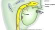
Introduction: The facial canal (FC) is an extensive bony canal that houses the facial nerve and occupies a central position in the petrous part of temporal bone. It is of utmost significance to otologists due to its dehiscence and relationship to the inner or middle ear components. The main objectives of current investigation are to detect variations in the reported values of FC anatomy that may occur due to different methodology and to elucidate the influence of age and ethnic factors on the morphological features of FC.
Methods: The methodology is adapted to the Preferred Reporting Items for Systematic Reviews and Meta-Analyses (PRISMA) guidelines. Pooled weighted estimation was performed to calculate the mean length, angle, and prevalence of dehiscence.
Results: The cross-sectional shape of FC varied from circular to ellipsoid index and is 1.45 [95% CI, 0.86-2.6]. The mean length of the FC is 34.42 mm [95% CI, 27.62-40.13 mm] and the mean width or diameter is 1.35 mm [95% CI, 1.013-1.63 mm]. The length of the FC in fetuses and children is 21.79 mm [95% CI, 18.44-25.15 mm], and 26.92 mm [95% CI, 23.3-28.3 mm], respectively. In meta-regression, age is observed as a predictor and accounts for 36% of the heterogeneity. The prevalence of FC dehiscence in healthy temporal bones is 29% [95% CI, 20-40%].
Conclusion: The different segments of the FC exhibit significant variability and an unusually high incidence of dehiscence, which could potentially have clinical implications for the etiopathogenesis of facial nerve dysfunction.
期刊介绍:
Anatomy is a morphological science which cannot fail to interest the clinician. The practical application of anatomical research to clinical problems necessitates special adaptation and selectivity in choosing from numerous international works. Although there is a tendency to believe that meaningful advances in anatomy are unlikely, constant revision is necessary. Surgical and Radiologic Anatomy, the first international journal of Clinical anatomy has been created in this spirit.
Its goal is to serve clinicians, regardless of speciality-physicians, surgeons, radiologists or other specialists-as an indispensable aid with which they can improve their knowledge of anatomy. Each issue includes: Original papers, review articles, articles on the anatomical bases of medical, surgical and radiological techniques, articles of normal radiologic anatomy, brief reviews of anatomical publications of clinical interest.
Particular attention is given to high quality illustrations, which are indispensable for a better understanding of anatomical problems.
Surgical and Radiologic Anatomy is a journal written by anatomists for clinicians with a special interest in anatomy.

 求助内容:
求助内容: 应助结果提醒方式:


