{"title":"Ontogeny of the masticatory muscles in the opossum Didelphis albiventris (Marsupialia, Didelphimorphia, Didelphidae)","authors":"Juann A. F. H. Abreu, Diego Astúa","doi":"10.1111/joa.14109","DOIUrl":null,"url":null,"abstract":"<p>Opossums (marsupials of the Didelphidae family) retain a generalized masticatory apparatus and tribosphenic molars, often used as models to understand the evolution of mastication in early therian mammals. Like all marsupials, their growth goes through a stage when pups complete their development while permanently attached to the mother's teats before weaning and starting feeding on their own. Yet, while the masticatory muscles of adults are known, as is the ontogeny of the cranium and mandible, the ontogenetic changes in the masticatory muscles remain unknown. Here we describe for the first time the changes in the masticatory muscles observed in lactating pups, and weaned juveniles, subadults, and adults in the White-eared opossum, <i>Didelphis albiventris</i>, through dissection of 25 specimens and quantification of relative muscle masses, lines of actions and mechanical advantages whenever possible. We also assessed the scaling patterns of muscle masses and mechanical advantages through ontogeny. The main changes, as expected, were found between suckling and weaned specimens, although some changes still occurred from juveniles to adults. The adult adductor musculature is similar to the other <i>Didelphis</i> species already known, with a dominant <i>m. temporalis</i> that originates on the lateral wall of the skull, up to the sagittal and nuchal crests, and fills the zygomatic arch when inserting into the lateral and medial surfaces of the coronoid process, respectively through the <i>pars superficialis</i> and <i>pars profunda.</i> The <i>m. masseter</i> is also subdivided in superficial and deep bundles which originate posteriorly in the maxilla and zygomatic arch, and insert into the angular process and masseteric fossa in the mandible. The <i>m. pterygoideus medialis</i> originates from the palatine, the pterygoid bone and the alisphenoid, and it inserts on the angular process medially. Suckling pups showed muscles with more restricted attachments, reduced muscle lines of action, and less diversity in the fiber orientation. The absence of the postorbital constriction also resulted in a distinct morphology of the <i>m. temporalis pars profunda</i>, through two bundles, one anterior and one posterior, which insert more inferiorly into the mandible. These major changes can be related to the onset of mastication and to size-related changes in growing weaned age classes. In general, all adductor muscles grew with positive allometry, and increased their fixation areas through, in part, the development of specific regions of the cranium and mandible. Their lines of action also increase and diversify along ontogeny. These changes can be related to the functional requirements for fixation during lactation, which shift to adduction and mastication movements after weaning.</p>","PeriodicalId":14971,"journal":{"name":"Journal of Anatomy","volume":"245 4","pages":"625-642"},"PeriodicalIF":1.8000,"publicationDate":"2024-07-12","publicationTypes":"Journal Article","fieldsOfStudy":null,"isOpenAccess":false,"openAccessPdf":"","citationCount":"0","resultStr":null,"platform":"Semanticscholar","paperid":null,"PeriodicalName":"Journal of Anatomy","FirstCategoryId":"3","ListUrlMain":"https://onlinelibrary.wiley.com/doi/10.1111/joa.14109","RegionNum":3,"RegionCategory":"医学","ArticlePicture":[],"TitleCN":null,"AbstractTextCN":null,"PMCID":null,"EPubDate":"","PubModel":"","JCR":"Q2","JCRName":"ANATOMY & MORPHOLOGY","Score":null,"Total":0}
引用次数: 0
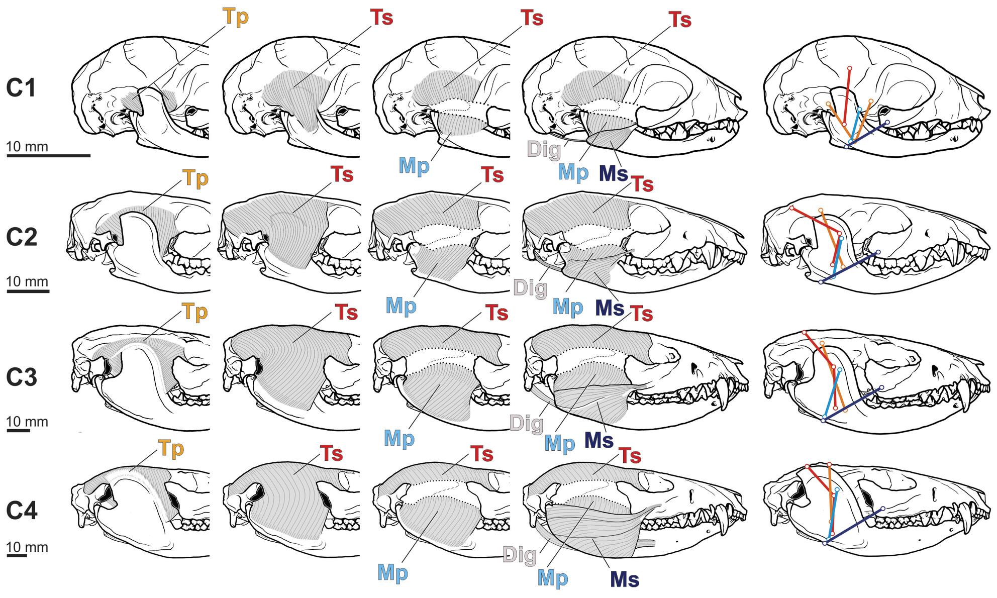
Abstract
Opossums (marsupials of the Didelphidae family) retain a generalized masticatory apparatus and tribosphenic molars, often used as models to understand the evolution of mastication in early therian mammals. Like all marsupials, their growth goes through a stage when pups complete their development while permanently attached to the mother's teats before weaning and starting feeding on their own. Yet, while the masticatory muscles of adults are known, as is the ontogeny of the cranium and mandible, the ontogenetic changes in the masticatory muscles remain unknown. Here we describe for the first time the changes in the masticatory muscles observed in lactating pups, and weaned juveniles, subadults, and adults in the White-eared opossum, Didelphis albiventris, through dissection of 25 specimens and quantification of relative muscle masses, lines of actions and mechanical advantages whenever possible. We also assessed the scaling patterns of muscle masses and mechanical advantages through ontogeny. The main changes, as expected, were found between suckling and weaned specimens, although some changes still occurred from juveniles to adults. The adult adductor musculature is similar to the other Didelphis species already known, with a dominant m. temporalis that originates on the lateral wall of the skull, up to the sagittal and nuchal crests, and fills the zygomatic arch when inserting into the lateral and medial surfaces of the coronoid process, respectively through the pars superficialis and pars profunda. The m. masseter is also subdivided in superficial and deep bundles which originate posteriorly in the maxilla and zygomatic arch, and insert into the angular process and masseteric fossa in the mandible. The m. pterygoideus medialis originates from the palatine, the pterygoid bone and the alisphenoid, and it inserts on the angular process medially. Suckling pups showed muscles with more restricted attachments, reduced muscle lines of action, and less diversity in the fiber orientation. The absence of the postorbital constriction also resulted in a distinct morphology of the m. temporalis pars profunda, through two bundles, one anterior and one posterior, which insert more inferiorly into the mandible. These major changes can be related to the onset of mastication and to size-related changes in growing weaned age classes. In general, all adductor muscles grew with positive allometry, and increased their fixation areas through, in part, the development of specific regions of the cranium and mandible. Their lines of action also increase and diversify along ontogeny. These changes can be related to the functional requirements for fixation during lactation, which shift to adduction and mastication movements after weaning.
期刊介绍:
Journal of Anatomy is an international peer-reviewed journal sponsored by the Anatomical Society. The journal publishes original papers, invited review articles and book reviews. Its main focus is to understand anatomy through an analysis of structure, function, development and evolution. Priority will be given to studies of that clearly articulate their relevance to the anatomical community. Focal areas include: experimental studies, contributions based on molecular and cell biology and on the application of modern imaging techniques and papers with novel methods or synthetic perspective on an anatomical system.
Studies that are essentially descriptive anatomy are appropriate only if they communicate clearly a broader functional or evolutionary significance. You must clearly state the broader implications of your work in the abstract.
We particularly welcome submissions in the following areas:
Cell biology and tissue architecture
Comparative functional morphology
Developmental biology
Evolutionary developmental biology
Evolutionary morphology
Functional human anatomy
Integrative vertebrate paleontology
Methodological innovations in anatomical research
Musculoskeletal system
Neuroanatomy and neurodegeneration
Significant advances in anatomical education.

 求助内容:
求助内容: 应助结果提醒方式:


