{"title":"Ultra-high b-value DWI in rectal cancer: image quality assessment and regional lymph node prediction based on radiomics.","authors":"Yongfei Hao, Jianyong Zheng, Wanqing Li, Wanting Zhao, Jianmin Zheng, Hong Wang, Jialiang Ren, Guangwen Zhang, Jinsong Zhang","doi":"10.1007/s00330-024-10958-3","DOIUrl":null,"url":null,"abstract":"<p><strong>Objectives: </strong>This study aims to evaluate image quality and regional lymph node metastasis (LNM) in patients with rectal cancer (RC) on multi-b-value diffusion-weighted imaging (DWI).</p><p><strong>Methods: </strong>This retrospective study included 199 patients with RC who had undergone multi-b-value DWI. Subjective (five-point Likert scale) and objective assessments of quality images were performed on DWI<sub>b1000</sub>, DWI<sub>b2000,</sub> and DWI<sub>b3000</sub>. Patients were randomly divided into a training (n = 140) or validation cohort (n = 59). Radiomics features were extracted within the whole volume tumor on ADC maps (b = 0, 1000 s/mm<sup>2</sup>), DWI<sub>b1000</sub>, DWI<sub>b2000</sub>, and DWI<sub>b3000</sub>, respectively. Five prediction models based on selected features were developed using logistic regression analysis. The performance of radiomics models was evaluated with a receiver operating characteristic curve, calibration, and decision curve analysis (DCA).</p><p><strong>Results: </strong>The mean signal intensity of the tumor (SI<sub>tumor</sub>), signal-to-noise ratio (SNR), and artifact and anatomic differentiability score gradually were decreased as the b-value increased. However, the contrast-to-noise (CNR) on DWI<sub>b2000</sub> was superior to those of DWI<sub>b1000</sub> and DWI<sub>b3000</sub> (4.58 ± 0.86, 3.82 ± 0.77, 4.18 ± 0.84, p < 0.001, respectively). The overall image quality score of DWI<sub>b2000</sub> was higher than that of DWI<sub>b3000</sub> (p < 0.001) and showed no significant difference between DWI<sub>b1000</sub> and DWI<sub>b2000</sub> (p = 0.059). The area under curve (AUC) value of the radiomics model based on DWI<sub>b2000</sub> (0.728) was higher than conventional ADC maps (0.690), DWI<sub>b1000</sub> (0.699), and DWI<sub>b3000</sub> (0.707), but inferior to multi-b-value DWI (0.739) in predicting LNM.</p><p><strong>Conclusion: </strong>DWI<sub>b2000</sub> provides better lesion conspicuity and LNM prediction than DWI<sub>b1000</sub> and DWI<sub>b3000</sub> in RC.</p><p><strong>Clinical relevance statement: </strong>DWI<sub>b2000</sub> offers satisfactory visualization of lesions. Radiomics features based on DWI<sub>b2000</sub> can be applied for preoperatively predicting regional lymph node metastasis in rectal cancer, thereby benefiting the stratified treatment strategy.</p><p><strong>Key points: </strong>Lymph node staging is required to determine the best treatment plan for rectal cancer. DWI<sub>b2000</sub> provides superior contrast-to-noise ratio and lesion conspicuity and its derived radiomics best predict lymph node metastasis. DWI<sub>b2000</sub> may be recommended as the optimal b-value in rectal MRI protocol.</p>","PeriodicalId":12076,"journal":{"name":"European Radiology","volume":" ","pages":"49-60"},"PeriodicalIF":4.7000,"publicationDate":"2025-01-01","publicationTypes":"Journal Article","fieldsOfStudy":null,"isOpenAccess":false,"openAccessPdf":"","citationCount":"0","resultStr":null,"platform":"Semanticscholar","paperid":null,"PeriodicalName":"European Radiology","FirstCategoryId":"3","ListUrlMain":"https://doi.org/10.1007/s00330-024-10958-3","RegionNum":2,"RegionCategory":"医学","ArticlePicture":[],"TitleCN":null,"AbstractTextCN":null,"PMCID":null,"EPubDate":"2024/7/12 0:00:00","PubModel":"Epub","JCR":"Q1","JCRName":"RADIOLOGY, NUCLEAR MEDICINE & MEDICAL IMAGING","Score":null,"Total":0}
引用次数: 0
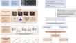
Abstract
Objectives: This study aims to evaluate image quality and regional lymph node metastasis (LNM) in patients with rectal cancer (RC) on multi-b-value diffusion-weighted imaging (DWI).
Methods: This retrospective study included 199 patients with RC who had undergone multi-b-value DWI. Subjective (five-point Likert scale) and objective assessments of quality images were performed on DWIb1000, DWIb2000, and DWIb3000. Patients were randomly divided into a training (n = 140) or validation cohort (n = 59). Radiomics features were extracted within the whole volume tumor on ADC maps (b = 0, 1000 s/mm2), DWIb1000, DWIb2000, and DWIb3000, respectively. Five prediction models based on selected features were developed using logistic regression analysis. The performance of radiomics models was evaluated with a receiver operating characteristic curve, calibration, and decision curve analysis (DCA).
Results: The mean signal intensity of the tumor (SItumor), signal-to-noise ratio (SNR), and artifact and anatomic differentiability score gradually were decreased as the b-value increased. However, the contrast-to-noise (CNR) on DWIb2000 was superior to those of DWIb1000 and DWIb3000 (4.58 ± 0.86, 3.82 ± 0.77, 4.18 ± 0.84, p < 0.001, respectively). The overall image quality score of DWIb2000 was higher than that of DWIb3000 (p < 0.001) and showed no significant difference between DWIb1000 and DWIb2000 (p = 0.059). The area under curve (AUC) value of the radiomics model based on DWIb2000 (0.728) was higher than conventional ADC maps (0.690), DWIb1000 (0.699), and DWIb3000 (0.707), but inferior to multi-b-value DWI (0.739) in predicting LNM.
Conclusion: DWIb2000 provides better lesion conspicuity and LNM prediction than DWIb1000 and DWIb3000 in RC.
Clinical relevance statement: DWIb2000 offers satisfactory visualization of lesions. Radiomics features based on DWIb2000 can be applied for preoperatively predicting regional lymph node metastasis in rectal cancer, thereby benefiting the stratified treatment strategy.
Key points: Lymph node staging is required to determine the best treatment plan for rectal cancer. DWIb2000 provides superior contrast-to-noise ratio and lesion conspicuity and its derived radiomics best predict lymph node metastasis. DWIb2000 may be recommended as the optimal b-value in rectal MRI protocol.
期刊介绍:
European Radiology (ER) continuously updates scientific knowledge in radiology by publication of strong original articles and state-of-the-art reviews written by leading radiologists. A well balanced combination of review articles, original papers, short communications from European radiological congresses and information on society matters makes ER an indispensable source for current information in this field.
This is the Journal of the European Society of Radiology, and the official journal of a number of societies.
From 2004-2008 supplements to European Radiology were published under its companion, European Radiology Supplements, ISSN 1613-3749.

 求助内容:
求助内容: 应助结果提醒方式:


