Lingui Gu, Hualin Chen, Mingjiang Sun, Yihao Chen, Qinglei Shi, Jianbo Chang, Junji Wei, Wenbin Ma, Xinjie Bao, Renzhi Wang
{"title":"Unraveling dynamic immunological landscapes in intracerebral hemorrhage: insights from single-cell and spatial transcriptomic profiling","authors":"Lingui Gu, Hualin Chen, Mingjiang Sun, Yihao Chen, Qinglei Shi, Jianbo Chang, Junji Wei, Wenbin Ma, Xinjie Bao, Renzhi Wang","doi":"10.1002/mco2.635","DOIUrl":null,"url":null,"abstract":"<p>Intracerebral hemorrhage (ICH) poses a formidable challenge in stroke management, with limited therapeutic options, particularly in the realm of immune-targeted interventions. Clinical trials targeting immune responses post-ICH have encountered setbacks, potentially attributable to the substantial cellular heterogeneity and intricate intercellular networks within the brain. Here, we present a pioneering investigation utilizing single-cell RNA sequencing and spatial transcriptome profiling at hyperacute (1 h), acute (24 h), and subacute (7 days) intervals post-ICH, aimed at unraveling the dynamic immunological landscape and spatial distributions within the cerebral tissue. Our comprehensive analysis revealed distinct cell differentiation patterns among myeloid and lymphocyte populations, along with delineated spatial distributions across various brain regions. Notably, we identified a subset of lymphocytes characterized by the expression of Spp1 and Lyz2, termed macrophage-associated lymphocytes, which exhibited close interactions with myeloid cells. Specifically, we observed prominent interactions between Lgmn+Macro-T cells and microglia through the spp1–cd44 pathway during the acute phase post-ICH in the choroid plexus. These findings represent a significant advancement in our understanding of immune cell dynamics at single-cell resolution across distinct post-ICH time points, thereby laying the groundwork for exploring critical temporal windows and informing the development of targeted therapeutic strategies.</p>","PeriodicalId":94133,"journal":{"name":"MedComm","volume":null,"pages":null},"PeriodicalIF":10.7000,"publicationDate":"2024-07-10","publicationTypes":"Journal Article","fieldsOfStudy":null,"isOpenAccess":false,"openAccessPdf":"https://www.ncbi.nlm.nih.gov/pmc/articles/PMC11233862/pdf/","citationCount":"0","resultStr":null,"platform":"Semanticscholar","paperid":null,"PeriodicalName":"MedComm","FirstCategoryId":"1085","ListUrlMain":"https://onlinelibrary.wiley.com/doi/10.1002/mco2.635","RegionNum":0,"RegionCategory":null,"ArticlePicture":[],"TitleCN":null,"AbstractTextCN":null,"PMCID":null,"EPubDate":"","PubModel":"","JCR":"Q1","JCRName":"MEDICINE, RESEARCH & EXPERIMENTAL","Score":null,"Total":0}
引用次数: 0
Abstract
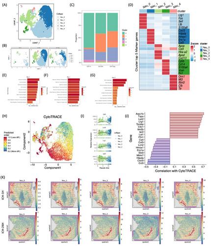
Intracerebral hemorrhage (ICH) poses a formidable challenge in stroke management, with limited therapeutic options, particularly in the realm of immune-targeted interventions. Clinical trials targeting immune responses post-ICH have encountered setbacks, potentially attributable to the substantial cellular heterogeneity and intricate intercellular networks within the brain. Here, we present a pioneering investigation utilizing single-cell RNA sequencing and spatial transcriptome profiling at hyperacute (1 h), acute (24 h), and subacute (7 days) intervals post-ICH, aimed at unraveling the dynamic immunological landscape and spatial distributions within the cerebral tissue. Our comprehensive analysis revealed distinct cell differentiation patterns among myeloid and lymphocyte populations, along with delineated spatial distributions across various brain regions. Notably, we identified a subset of lymphocytes characterized by the expression of Spp1 and Lyz2, termed macrophage-associated lymphocytes, which exhibited close interactions with myeloid cells. Specifically, we observed prominent interactions between Lgmn+Macro-T cells and microglia through the spp1–cd44 pathway during the acute phase post-ICH in the choroid plexus. These findings represent a significant advancement in our understanding of immune cell dynamics at single-cell resolution across distinct post-ICH time points, thereby laying the groundwork for exploring critical temporal windows and informing the development of targeted therapeutic strategies.

 求助内容:
求助内容: 应助结果提醒方式:


