{"title":"Measurement of human peritoneal surface area using artificial intelligence software in abdominal computed tomography.","authors":"Seung Joon Choi, Ji-Hyeon Park, Youngbae Jeon, Donghyuk Lee, Jeong-Heum Baek","doi":"10.14216/kjco.24002","DOIUrl":null,"url":null,"abstract":"<p><strong>Purpose: </strong>The calculation of the intraperitoneal organ surface area is important for understanding their anatomical structure and for conducting basic and clinical studies on diseases related to the peritoneum. To measure the intraperitoneal surface area in a living body by applying artificial intelligence (AI) techniques to the abdominal cavity using computed tomography and to prepare clinical indicators for application to the abdominal cavity.</p><p><strong>Methods: </strong>Computed tomography images of ten adult males and females with a healthy body mass index and ten adults diagnosed with colon cancer were analyzed to determine the peritoneal and intraperitoneal surface areas of the organs. The peritoneal surface was segmented and three-dimensionally modeled using AI medical imaging software. In addition to manual work, three-dimensional editing, filtering, and connectivity checks were performed to improve work efficiency and accuracy. The colon and small intestine surface areas were calculated using the mean length and diameter. The abdominal cavity surface area was defined as the sum of the intraperitoneal area and the surface areas of each organ.</p><p><strong>Results: </strong>The mean peritoneal surface area of all participants was measured as 10,039 ± 241 cm2 (males 10,224 ± 171 cm2 and females 9,854 ± 134 cm2). Males had a 3.7% larger peritoneal surface area than females, with a statistically significant difference (P < 0.001).</p><p><strong>Conclusion: </strong>The abdominal cavity surface area can be measured using AI techniques and is expected to be used as basic data for clinical applications.</p>","PeriodicalId":74045,"journal":{"name":"Korean journal of clinical oncology","volume":"20 1","pages":"6-12"},"PeriodicalIF":0.0000,"publicationDate":"2024-05-01","publicationTypes":"Journal Article","fieldsOfStudy":null,"isOpenAccess":false,"openAccessPdf":"https://www.ncbi.nlm.nih.gov/pmc/articles/PMC11261175/pdf/","citationCount":"0","resultStr":null,"platform":"Semanticscholar","paperid":null,"PeriodicalName":"Korean journal of clinical oncology","FirstCategoryId":"1085","ListUrlMain":"https://doi.org/10.14216/kjco.24002","RegionNum":0,"RegionCategory":null,"ArticlePicture":[],"TitleCN":null,"AbstractTextCN":null,"PMCID":null,"EPubDate":"2024/6/30 0:00:00","PubModel":"Epub","JCR":"","JCRName":"","Score":null,"Total":0}
引用次数: 0
Abstract
Purpose: The calculation of the intraperitoneal organ surface area is important for understanding their anatomical structure and for conducting basic and clinical studies on diseases related to the peritoneum. To measure the intraperitoneal surface area in a living body by applying artificial intelligence (AI) techniques to the abdominal cavity using computed tomography and to prepare clinical indicators for application to the abdominal cavity.
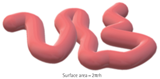
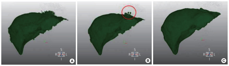
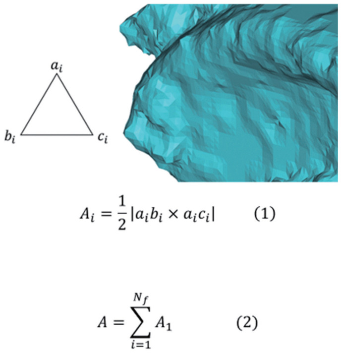
Methods: Computed tomography images of ten adult males and females with a healthy body mass index and ten adults diagnosed with colon cancer were analyzed to determine the peritoneal and intraperitoneal surface areas of the organs. The peritoneal surface was segmented and three-dimensionally modeled using AI medical imaging software. In addition to manual work, three-dimensional editing, filtering, and connectivity checks were performed to improve work efficiency and accuracy. The colon and small intestine surface areas were calculated using the mean length and diameter. The abdominal cavity surface area was defined as the sum of the intraperitoneal area and the surface areas of each organ.
Results: The mean peritoneal surface area of all participants was measured as 10,039 ± 241 cm2 (males 10,224 ± 171 cm2 and females 9,854 ± 134 cm2). Males had a 3.7% larger peritoneal surface area than females, with a statistically significant difference (P < 0.001).
Conclusion: The abdominal cavity surface area can be measured using AI techniques and is expected to be used as basic data for clinical applications.

 求助内容:
求助内容: 应助结果提醒方式:


