Theo Chenal, Fanny Granat, Catherine Trumel, Nathalie Bourgès-Abella
{"title":"What is your diagnosis? Abnormal cluster on the WDF and WNR scattergrams from Sysmex XN-V in a dog","authors":"Theo Chenal, Fanny Granat, Catherine Trumel, Nathalie Bourgès-Abella","doi":"10.1111/vcp.13362","DOIUrl":null,"url":null,"abstract":"<p>A 4-month-old intact male, Australian shepherd dog was presented to the emergency unit of the veterinary school hospital of Toulouse (France) for the medical care of a canine Parvovirus infection diagnosed from a positive snap test (SNAP Parvo, IDEXX Laboratories, Westbrook, USA). Clinical examination revealed pale mucous membranes and palpable fluid in the abdomen.</p><p>A CBC performed with the ProCyte Dx (IDEXX, Westbrook, USA) (Table S1) revealed a marked normocytic normochromic poorly regenerative anemia, mild leukocytosis, neutrophilia, monocytosis, and marked thrombocytopenia with a flag. The thrombocytopenia was suspected to be genuine despite the observation of a few platelet-fibrin clots on the blood smear. An abdominal ultrasound confirmed the presence of a peritoneal effusion. The dog was transfused with compatible blood.</p><p>The following day, analysis of the blood and effusion was performed with the Sysmex XN-V (Sysmex, Kobe, Japan) (Table S1), and smears were reviewed. The CBC confirmed the anemia and revealed mild neutrophilia and lymphopenia with an abnormal WDF scattergram (Figure 1). A marked thrombocytopenia with a flag was reported and confirmed by the absence of platelet clumps on blood smear and a manual estimate of platelet count (12.10<sup>9</sup>/L). The effusion was macroscopically bloody, results were very similar to the CBC (Table S1), and the cytologic appearance confirmed the suspicion of hemoperitoneum, possibly secondary to the thrombocytopenia even though no bruising or petechia were observed; the hemostasis panel (thrombin time, activated partial thromboplastin time and fibrinogen concentration) was within reference interval.</p><p>During hospitalization, two additional CBCs performed with the Sysmex XN-V revealed no abnormal cluster.</p><p>To confirm our hypothesis, we added 1 μL of ultrasound gel (Supragel, LCH medical product, Paris, France) in 1 mL of fresh canine EDTA-blood specimen using leftover blood from a blood donation.</p><p>A CBC and smear examination of both the native specimen and the specimen mixed with the gel were performed. CBC of the native blood specimen revealed mild erythrocytosis, and examination of the blood smear only revealed a few small platelet aggregates. A curvilinear cluster very similar to the one described in our case was observed on both WDF and WNR scattergrams (Figure 4) with the specimen mixed with gel, resulting in a slight overestimation of WBC and eosinophil counts. Other scattergrams were normal. On the smear, granular material similar to the one observed in our case was also visualized.</p><p>This case reports an unusual CBC finding characterized by an additional curvilinear cluster on WDF and WNR scattergrams obtained with the Sysmex XN-V analyzer secondary to blood contamination with ultrasound gel. To our knowledge, there are only a few reports of unusual Sysmex XN-V WDF and WNR scattergrams in human and veterinary medicine.</p><p>A curvilinear pattern on the WDF scattergram was observed using the Sysmex XN-1000 in body fluid mode in a cerebrospinal fluid from a patient with lymphoma metastasized to the brain and treated with intrathecal DepoCyt, a drug composed of cytarabine in liposomal particles.<span><sup>1</sup></span> Authors suggested, based on experiments conducted on blood mixed with DepoCyt, that liposomal particles were similar in size, had similar physical properties to WBCs, and fell into similar areas of WDF scattergram, leading to an overestimation of cell count.</p><p>Another curvilinear cluster was observed using the Sysmex XN-9000 with blood specimens from two human patients with circulating giant platelets and megakaryocytes secondary to type 2-refractory anemia with excess blasts.<span><sup>2</sup></span></p><p>Layssol-Lamour et al also reported a curvilinear interference using the ProCyte Dx in mice and rats with prominent platelet aggregation.<span><sup>3</sup></span> This has been routinely observed by the authors with the Sysmex XN-V and is usually more prominent in cats and goats than in dogs but never as pronounced as in this case (personal observation). In the validation study of the Sysmex XN-V in dogs, no interference was described with platelet aggregation, but the presence of abnormal leukocytes (band cells and acute leukemias) led to modifications of WNR and/or WDF scattergrams characterized by fusion of clusters and arbitrary separation of populations on the WDF scattergram due to the presence of immature cells (often misclassified as monocytes and lymphocytes), rather than an additional curvilinear cluster originating from debris and unlysed RBC.<span><sup>4</sup></span></p><p>In our case, similar findings with experimental contamination confirmed that the interference was secondary to the ultrasound gel. Some ultrasound gel material has clearly been associated with artifactual images on slides stained with Romanowsky stains.<span><sup>5</sup></span> It would be interesting to know if all gel materials cause interference with the Sysmex XN-V scattergrams and WBC counts. As the blood was collected following an ultrasound examination and given that even a small amount of gel can cause substantial interference, contamination of equipment (i.e., gloves used because of the “infectious” status of the patient) is suspected to have caused the blood contamination, probably by contaminating the sampling area or material.</p><p>This article describes a report of an additional curvilinear cluster on the WDF and WNR scattergrams of the Sysmex XN-V analyzer secondary to contamination of canine blood with ultrasound gel.</p><p>It emphasizes the fact that in the case of abnormal clusters on WBC scattergrams, a blood smear examination is mandatory to validate analyzer results. Furthermore, it highlights the importance of properly clearing ultrasound gel before sampling a biological fluid to avoid potential interferences and spuriously high WBC counts when using the Sysmex XN-V.</p><p>The Sysmex XN-V is on loan from Sysmex.</p>","PeriodicalId":23593,"journal":{"name":"Veterinary clinical pathology","volume":"54 S1","pages":"S5-S8"},"PeriodicalIF":1.1000,"publicationDate":"2024-07-10","publicationTypes":"Journal Article","fieldsOfStudy":null,"isOpenAccess":false,"openAccessPdf":"https://onlinelibrary.wiley.com/doi/epdf/10.1111/vcp.13362","citationCount":"0","resultStr":null,"platform":"Semanticscholar","paperid":null,"PeriodicalName":"Veterinary clinical pathology","FirstCategoryId":"97","ListUrlMain":"https://onlinelibrary.wiley.com/doi/10.1111/vcp.13362","RegionNum":4,"RegionCategory":"农林科学","ArticlePicture":[],"TitleCN":null,"AbstractTextCN":null,"PMCID":null,"EPubDate":"","PubModel":"","JCR":"Q3","JCRName":"VETERINARY SCIENCES","Score":null,"Total":0}
引用次数: 0
Abstract
A 4-month-old intact male, Australian shepherd dog was presented to the emergency unit of the veterinary school hospital of Toulouse (France) for the medical care of a canine Parvovirus infection diagnosed from a positive snap test (SNAP Parvo, IDEXX Laboratories, Westbrook, USA). Clinical examination revealed pale mucous membranes and palpable fluid in the abdomen.
A CBC performed with the ProCyte Dx (IDEXX, Westbrook, USA) (Table S1) revealed a marked normocytic normochromic poorly regenerative anemia, mild leukocytosis, neutrophilia, monocytosis, and marked thrombocytopenia with a flag. The thrombocytopenia was suspected to be genuine despite the observation of a few platelet-fibrin clots on the blood smear. An abdominal ultrasound confirmed the presence of a peritoneal effusion. The dog was transfused with compatible blood.
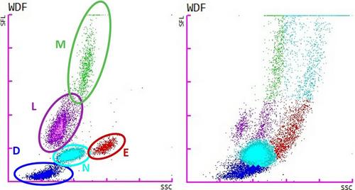
The following day, analysis of the blood and effusion was performed with the Sysmex XN-V (Sysmex, Kobe, Japan) (Table S1), and smears were reviewed. The CBC confirmed the anemia and revealed mild neutrophilia and lymphopenia with an abnormal WDF scattergram (Figure 1). A marked thrombocytopenia with a flag was reported and confirmed by the absence of platelet clumps on blood smear and a manual estimate of platelet count (12.109/L). The effusion was macroscopically bloody, results were very similar to the CBC (Table S1), and the cytologic appearance confirmed the suspicion of hemoperitoneum, possibly secondary to the thrombocytopenia even though no bruising or petechia were observed; the hemostasis panel (thrombin time, activated partial thromboplastin time and fibrinogen concentration) was within reference interval.
During hospitalization, two additional CBCs performed with the Sysmex XN-V revealed no abnormal cluster.
To confirm our hypothesis, we added 1 μL of ultrasound gel (Supragel, LCH medical product, Paris, France) in 1 mL of fresh canine EDTA-blood specimen using leftover blood from a blood donation.
A CBC and smear examination of both the native specimen and the specimen mixed with the gel were performed. CBC of the native blood specimen revealed mild erythrocytosis, and examination of the blood smear only revealed a few small platelet aggregates. A curvilinear cluster very similar to the one described in our case was observed on both WDF and WNR scattergrams (Figure 4) with the specimen mixed with gel, resulting in a slight overestimation of WBC and eosinophil counts. Other scattergrams were normal. On the smear, granular material similar to the one observed in our case was also visualized.
This case reports an unusual CBC finding characterized by an additional curvilinear cluster on WDF and WNR scattergrams obtained with the Sysmex XN-V analyzer secondary to blood contamination with ultrasound gel. To our knowledge, there are only a few reports of unusual Sysmex XN-V WDF and WNR scattergrams in human and veterinary medicine.
A curvilinear pattern on the WDF scattergram was observed using the Sysmex XN-1000 in body fluid mode in a cerebrospinal fluid from a patient with lymphoma metastasized to the brain and treated with intrathecal DepoCyt, a drug composed of cytarabine in liposomal particles.1 Authors suggested, based on experiments conducted on blood mixed with DepoCyt, that liposomal particles were similar in size, had similar physical properties to WBCs, and fell into similar areas of WDF scattergram, leading to an overestimation of cell count.
Another curvilinear cluster was observed using the Sysmex XN-9000 with blood specimens from two human patients with circulating giant platelets and megakaryocytes secondary to type 2-refractory anemia with excess blasts.2
Layssol-Lamour et al also reported a curvilinear interference using the ProCyte Dx in mice and rats with prominent platelet aggregation.3 This has been routinely observed by the authors with the Sysmex XN-V and is usually more prominent in cats and goats than in dogs but never as pronounced as in this case (personal observation). In the validation study of the Sysmex XN-V in dogs, no interference was described with platelet aggregation, but the presence of abnormal leukocytes (band cells and acute leukemias) led to modifications of WNR and/or WDF scattergrams characterized by fusion of clusters and arbitrary separation of populations on the WDF scattergram due to the presence of immature cells (often misclassified as monocytes and lymphocytes), rather than an additional curvilinear cluster originating from debris and unlysed RBC.4
In our case, similar findings with experimental contamination confirmed that the interference was secondary to the ultrasound gel. Some ultrasound gel material has clearly been associated with artifactual images on slides stained with Romanowsky stains.5 It would be interesting to know if all gel materials cause interference with the Sysmex XN-V scattergrams and WBC counts. As the blood was collected following an ultrasound examination and given that even a small amount of gel can cause substantial interference, contamination of equipment (i.e., gloves used because of the “infectious” status of the patient) is suspected to have caused the blood contamination, probably by contaminating the sampling area or material.
This article describes a report of an additional curvilinear cluster on the WDF and WNR scattergrams of the Sysmex XN-V analyzer secondary to contamination of canine blood with ultrasound gel.
It emphasizes the fact that in the case of abnormal clusters on WBC scattergrams, a blood smear examination is mandatory to validate analyzer results. Furthermore, it highlights the importance of properly clearing ultrasound gel before sampling a biological fluid to avoid potential interferences and spuriously high WBC counts when using the Sysmex XN-V.
期刊介绍:
Veterinary Clinical Pathology is the official journal of the American Society for Veterinary Clinical Pathology (ASVCP) and the European Society of Veterinary Clinical Pathology (ESVCP). The journal''s mission is to provide an international forum for communication and discussion of scientific investigations and new developments that advance the art and science of laboratory diagnosis in animals. Veterinary Clinical Pathology welcomes original experimental research and clinical contributions involving domestic, laboratory, avian, and wildlife species in the areas of hematology, hemostasis, immunopathology, clinical chemistry, cytopathology, surgical pathology, toxicology, endocrinology, laboratory and analytical techniques, instrumentation, quality assurance, and clinical pathology education.
 求助内容:
求助内容: 应助结果提醒方式:


