{"title":"Dynamic ultrasound assessment for insertional achilles tendinopathy: the COcco-RIcci (CORI) sign.","authors":"Giulio Cocco, Vincenzo Ricci","doi":"10.1007/s00256-024-04746-9","DOIUrl":null,"url":null,"abstract":"<p><strong>Objective: </strong>To describe a novel, dynamic ultrasound assessment of the Achilles tendon at the calcaneal insertion taking advantage of the effusion within the deep retrocalcaneal bursa as a natural contrast agent.</p><p><strong>Materials and methods: </strong>Positioning the ultrasound transducer in a longitudinal plane over the Achilles tendon at the calcaneal insertion, manual compression of the deep retrocalcaneal bursa can be performed using the other hand. Dynamically shifting the anechoic effusion from the proximal to the distal compartment of the bursa, the undersurface of the Achilles tendon is lifted from the underlying cortical bone of the superior facet of the calcaneal tuberosity.</p><p><strong>Results: </strong>Pushing the anechoic effusion from the bursal cavity toward the undersurface of the Achilles tendon, an eventual focal injury of its deep fibers can be visualized dynamically during the maneuver as a \"black crescent\" within the tendon-i.e., the COcco-RIcci (CORI) sign. Otherwise, the gliding of the posteroinferior wedge of the Kager's fat pad inside the tendon-bone interface can be observed in normal conditions.</p><p><strong>Conclusion: </strong>The CORI sign is a novel sonographic sign to further enhance the diagnostic accuracy of dynamic ultrasound imaging in patients with insertional Achilles tendinopathy especially to detect focal injury involving the deep fibers of the tendon.</p>","PeriodicalId":21783,"journal":{"name":"Skeletal Radiology","volume":" ","pages":"593-599"},"PeriodicalIF":1.9000,"publicationDate":"2025-03-01","publicationTypes":"Journal Article","fieldsOfStudy":null,"isOpenAccess":false,"openAccessPdf":"","citationCount":"0","resultStr":null,"platform":"Semanticscholar","paperid":null,"PeriodicalName":"Skeletal Radiology","FirstCategoryId":"3","ListUrlMain":"https://doi.org/10.1007/s00256-024-04746-9","RegionNum":3,"RegionCategory":"医学","ArticlePicture":[],"TitleCN":null,"AbstractTextCN":null,"PMCID":null,"EPubDate":"2024/7/10 0:00:00","PubModel":"Epub","JCR":"Q2","JCRName":"ORTHOPEDICS","Score":null,"Total":0}
引用次数: 0
Abstract
Objective: To describe a novel, dynamic ultrasound assessment of the Achilles tendon at the calcaneal insertion taking advantage of the effusion within the deep retrocalcaneal bursa as a natural contrast agent.
Materials and methods: Positioning the ultrasound transducer in a longitudinal plane over the Achilles tendon at the calcaneal insertion, manual compression of the deep retrocalcaneal bursa can be performed using the other hand. Dynamically shifting the anechoic effusion from the proximal to the distal compartment of the bursa, the undersurface of the Achilles tendon is lifted from the underlying cortical bone of the superior facet of the calcaneal tuberosity.
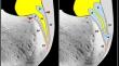
Results: Pushing the anechoic effusion from the bursal cavity toward the undersurface of the Achilles tendon, an eventual focal injury of its deep fibers can be visualized dynamically during the maneuver as a "black crescent" within the tendon-i.e., the COcco-RIcci (CORI) sign. Otherwise, the gliding of the posteroinferior wedge of the Kager's fat pad inside the tendon-bone interface can be observed in normal conditions.
Conclusion: The CORI sign is a novel sonographic sign to further enhance the diagnostic accuracy of dynamic ultrasound imaging in patients with insertional Achilles tendinopathy especially to detect focal injury involving the deep fibers of the tendon.
期刊介绍:
Skeletal Radiology provides a forum for the dissemination of current knowledge and information dealing with disorders of the musculoskeletal system including the spine. While emphasizing the radiological aspects of the many varied skeletal abnormalities, the journal also adopts an interdisciplinary approach, reflecting the membership of the International Skeletal Society. Thus, the anatomical, pathological, physiological, clinical, metabolic and epidemiological aspects of the many entities affecting the skeleton receive appropriate consideration.
This is the Journal of the International Skeletal Society and the Official Journal of the Society of Skeletal Radiology and the Australasian Musculoskelelal Imaging Group.

 求助内容:
求助内容: 应助结果提醒方式:


