{"title":"[<sup>68</sup>Ga]Ga-labeled FAPI Conjugated with Gly-Pro Sequence for PET Imaging of Malignant Tumors.","authors":"Yuxiang Shang, Guojin Zhang, Xinchao Yao, Chaoquan Lai, Fanghu Wang, Baozhen Zeng, Entao Liu, Hui Yuan, Zhen Cheng, Lei Jiang","doi":"10.1007/s11307-024-01935-9","DOIUrl":null,"url":null,"abstract":"<p><strong>Purpose: </strong>To improve tumor uptake and prolong tumor retention, a novel fibroblast activation protein (FAP) ligand based on a quinoline-based FAP inhibitor (FAPI) conjugated with the Gly-Pro sequence and 1,4,7,10-tetraazacyclododecane-N,N',N″,N‴-tetraacetic acid (DOTA) was radiolabeled with [<sup>68</sup>Ga]GaCl<sub>3</sub> ([<sup>68</sup>Ga]Ga-DOTA-GPFAPI-04). Due to the tumor heterogeneity, this study aimed to further validate the preclinical value of [<sup>68</sup>Ga]Ga-DOTA-GPFAPI-04 PET imaging in tumor mice models with different FAP expression levels.</p><p><strong>Methods: </strong>[<sup>68</sup>Ga]Ga-DOTA-GPFAPI-04 was synthesized and its partition coefficient was measured. The stability of [<sup>68</sup>Ga]Ga-DOTA-GPFAPI-04 was tested in phosphate-buffered saline (PBS, pH 7.4) and fetal bovine serum (FBS). Small animal PET and semi-quantitative studies were conducted in Panc-1 and A549 xenograft tumor mice models compared with [<sup>68</sup>Ga]Ga-DOTA-FAPI-04. Immunofluorescent and immunohistochemical staining and western blot assay were performed to confirm FAP expression in xenograft tumors.</p><p><strong>Results: </strong>[<sup>68</sup>Ga]Ga-DOTA-GPFAPI-04 exhibited a radiochemical purity of > 99% and high stability in PBS and FBS. [<sup>68</sup>Ga]Ga-DOTA-GPFAPI-04 had higher hydrophilic property than [<sup>68</sup>Ga]Ga-DOTA-FAPI-04 (-4.09 ± 0.05 vs -3.45 ± 0.05). Small animal PET and semi-quantitative analysis revealed Panc-1 xenograft tumor displayed higher tumor uptake of [<sup>68</sup>Ga]Ga-DOTA-GPFAPI-04 and tumor-to-background ratios compared to A549 xenograft tumor, consistent with the results of immunofluorescence, immunohistochemistry, and western blot. Moreover, [<sup>68</sup>Ga]Ga-DOTA-GPFAPI-04 demonstrated higher tumor accumulation and longer tumor retention than [<sup>68</sup>Ga]Ga-DOTA-FAPI-04 in both Panc-1 and A549 xenograft tumors. Furthermore, the FAP-binding specificity of [<sup>68</sup>Ga]Ga-DOTA-GPFAPI-04 was confirmed in vivo by co-injection of unlabeled GPFAPI-04.</p><p><strong>Conclusion: </strong>[<sup>68</sup>Ga]Ga-DOTA-GPFAPI-04 showed more favorable in vivo tumor imaging and longer tumor retention compared to [<sup>68</sup>Ga]Ga-DOTA-FAPI-04, which has high potential to be a promising PET probe for detecting FAP-positive tumors.</p>","PeriodicalId":18760,"journal":{"name":"Molecular Imaging and Biology","volume":" ","pages":"729-737"},"PeriodicalIF":3.0000,"publicationDate":"2024-08-01","publicationTypes":"Journal Article","fieldsOfStudy":null,"isOpenAccess":false,"openAccessPdf":"","citationCount":"0","resultStr":null,"platform":"Semanticscholar","paperid":null,"PeriodicalName":"Molecular Imaging and Biology","FirstCategoryId":"3","ListUrlMain":"https://doi.org/10.1007/s11307-024-01935-9","RegionNum":4,"RegionCategory":"医学","ArticlePicture":[],"TitleCN":null,"AbstractTextCN":null,"PMCID":null,"EPubDate":"2024/7/10 0:00:00","PubModel":"Epub","JCR":"Q2","JCRName":"RADIOLOGY, NUCLEAR MEDICINE & MEDICAL IMAGING","Score":null,"Total":0}
引用次数: 0
Abstract
Purpose: To improve tumor uptake and prolong tumor retention, a novel fibroblast activation protein (FAP) ligand based on a quinoline-based FAP inhibitor (FAPI) conjugated with the Gly-Pro sequence and 1,4,7,10-tetraazacyclododecane-N,N',N″,N‴-tetraacetic acid (DOTA) was radiolabeled with [68Ga]GaCl3 ([68Ga]Ga-DOTA-GPFAPI-04). Due to the tumor heterogeneity, this study aimed to further validate the preclinical value of [68Ga]Ga-DOTA-GPFAPI-04 PET imaging in tumor mice models with different FAP expression levels.
Methods: [68Ga]Ga-DOTA-GPFAPI-04 was synthesized and its partition coefficient was measured. The stability of [68Ga]Ga-DOTA-GPFAPI-04 was tested in phosphate-buffered saline (PBS, pH 7.4) and fetal bovine serum (FBS). Small animal PET and semi-quantitative studies were conducted in Panc-1 and A549 xenograft tumor mice models compared with [68Ga]Ga-DOTA-FAPI-04. Immunofluorescent and immunohistochemical staining and western blot assay were performed to confirm FAP expression in xenograft tumors.
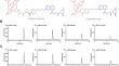
Results: [68Ga]Ga-DOTA-GPFAPI-04 exhibited a radiochemical purity of > 99% and high stability in PBS and FBS. [68Ga]Ga-DOTA-GPFAPI-04 had higher hydrophilic property than [68Ga]Ga-DOTA-FAPI-04 (-4.09 ± 0.05 vs -3.45 ± 0.05). Small animal PET and semi-quantitative analysis revealed Panc-1 xenograft tumor displayed higher tumor uptake of [68Ga]Ga-DOTA-GPFAPI-04 and tumor-to-background ratios compared to A549 xenograft tumor, consistent with the results of immunofluorescence, immunohistochemistry, and western blot. Moreover, [68Ga]Ga-DOTA-GPFAPI-04 demonstrated higher tumor accumulation and longer tumor retention than [68Ga]Ga-DOTA-FAPI-04 in both Panc-1 and A549 xenograft tumors. Furthermore, the FAP-binding specificity of [68Ga]Ga-DOTA-GPFAPI-04 was confirmed in vivo by co-injection of unlabeled GPFAPI-04.
Conclusion: [68Ga]Ga-DOTA-GPFAPI-04 showed more favorable in vivo tumor imaging and longer tumor retention compared to [68Ga]Ga-DOTA-FAPI-04, which has high potential to be a promising PET probe for detecting FAP-positive tumors.
期刊介绍:
Molecular Imaging and Biology (MIB) invites original contributions (research articles, review articles, commentaries, etc.) on the utilization of molecular imaging (i.e., nuclear imaging, optical imaging, autoradiography and pathology, MRI, MPI, ultrasound imaging, radiomics/genomics etc.) to investigate questions related to biology and health. The objective of MIB is to provide a forum to the discovery of molecular mechanisms of disease through the use of imaging techniques. We aim to investigate the biological nature of disease in patients and establish new molecular imaging diagnostic and therapy procedures.
Some areas that are covered are:
Preclinical and clinical imaging of macromolecular targets (e.g., genes, receptors, enzymes) involved in significant biological processes.
The design, characterization, and study of new molecular imaging probes and contrast agents for the functional interrogation of macromolecular targets.
Development and evaluation of imaging systems including instrumentation, image reconstruction algorithms, image analysis, and display.
Development of molecular assay approaches leading to quantification of the biological information obtained in molecular imaging.
Study of in vivo animal models of disease for the development of new molecular diagnostics and therapeutics.
Extension of in vitro and in vivo discoveries using disease models, into well designed clinical research investigations.
Clinical molecular imaging involving clinical investigations, clinical trials and medical management or cost-effectiveness studies.

 求助内容:
求助内容: 应助结果提醒方式:


