Pietro Arnaldi, Elena Casarotto, Michela Relucenti, Grazia Bellese, Maria Cristina Gagliani, Valeria Crippa, Patrizio Castagnola, Katia Cortese
{"title":"A NSC-34 cell line-derived spheroid model: Potential and challenges for in vitro evaluation of neurodegeneration","authors":"Pietro Arnaldi, Elena Casarotto, Michela Relucenti, Grazia Bellese, Maria Cristina Gagliani, Valeria Crippa, Patrizio Castagnola, Katia Cortese","doi":"10.1002/jemt.24651","DOIUrl":null,"url":null,"abstract":"<div>\n \n \n <section>\n \n <p>Three-dimensional (3D) spheroid models aim to bridge the gap between traditional two-dimensional (2D) cultures and the complex in vivo tissue environment. These models, created by self-clustering cells to mimic a 3D environment with surrounding extracellular framework, provide a valuable research tool. The NSC-34 cell line, generated by fusing mouse spinal cord motor neurons and neuroblastoma cells, is essential for studying neurodegenerative diseases like amyotrophic lateral sclerosis (ALS), where abnormal protein accumulation, such as TAR-DNA-binding protein 43 (TDP-43), occurs in affected nerve cells. However, NSC-34 behavior in a 3D context remains underexplored, and this study represents the first attempt to create a 3D model to determine its suitability for studying pathology. We generated NSC-34 spheroids using a nonadhesive hydrogel-based template and characterized them for 6 days. Light microscopy revealed that NSC-34 cells in 3D maintained high viability, a distinct round shape, and forming stable membrane connections. Scanning electron microscopy identified multiple tunnel-like structures, while ultrastructural analysis highlighted nuclear bending and mitochondria alterations. Using inducible GFP-TDP-43-expressing NSC-34 spheroids, we explored whether 3D structure affected TDP-43 expression, localization, and aggregation. Spheroids displayed nuclear GFP-TDP-43 expression, albeit at a reduced level compared with 2D cultures and generated both TDP-35 fragments and TDP-43 aggregates. This study sheds light on the distinctive behavior of NSC-34 in 3D culture, suggesting caution in the use of the 3D model for ALS or TDP-43 pathologies. Yet, it underscores the spheroids' potential for investigating fundamental cellular mechanisms, cell adaptation in a 3D context, future bioreactor applications, and drug penetration studies.</p>\n </section>\n \n <section>\n \n <h3> Research Highlights</h3>\n \n <div>\n <ul>\n \n <li>3D spheroid generation: NSC-34 spheroids, developed using a hydrogel-based template, showed high viability and distinct shapes for 6 days.</li>\n \n <li>Structural features: advanced microscopy identified tunnel-like structures and nuclear and mitochondrial changes in the spheroids.</li>\n \n <li>Protein dynamics: the study observed how 3D structures impact TDP-43 behavior, with altered expression but similar aggregation patterns to 2D cultures.</li>\n \n <li>Research implications: this study reveals the unique behavior of NSC-34 in 3D culture, suggests a careful approach to use this model for ALS or TDP-43 pathologies, and highlights its potential in cellular mechanism research and drug testing applications.</li>\n </ul>\n </div>\n </section>\n </div>","PeriodicalId":18684,"journal":{"name":"Microscopy Research and Technique","volume":null,"pages":null},"PeriodicalIF":2.0000,"publicationDate":"2024-07-11","publicationTypes":"Journal Article","fieldsOfStudy":null,"isOpenAccess":false,"openAccessPdf":"","citationCount":"0","resultStr":null,"platform":"Semanticscholar","paperid":null,"PeriodicalName":"Microscopy Research and Technique","FirstCategoryId":"5","ListUrlMain":"https://onlinelibrary.wiley.com/doi/10.1002/jemt.24651","RegionNum":3,"RegionCategory":"工程技术","ArticlePicture":[],"TitleCN":null,"AbstractTextCN":null,"PMCID":null,"EPubDate":"","PubModel":"","JCR":"Q2","JCRName":"ANATOMY & MORPHOLOGY","Score":null,"Total":0}
引用次数: 0
Abstract
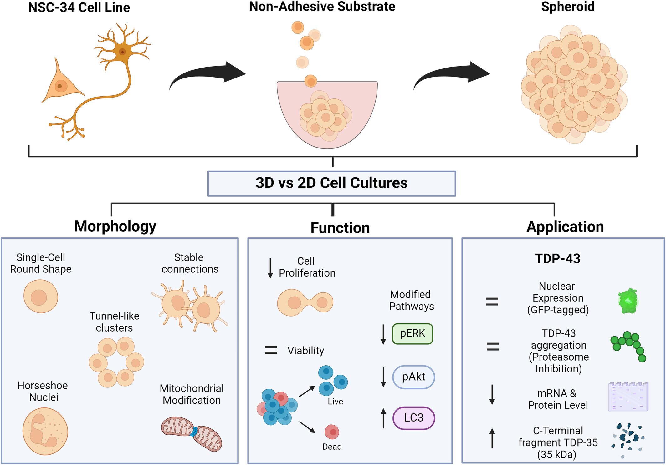
Three-dimensional (3D) spheroid models aim to bridge the gap between traditional two-dimensional (2D) cultures and the complex in vivo tissue environment. These models, created by self-clustering cells to mimic a 3D environment with surrounding extracellular framework, provide a valuable research tool. The NSC-34 cell line, generated by fusing mouse spinal cord motor neurons and neuroblastoma cells, is essential for studying neurodegenerative diseases like amyotrophic lateral sclerosis (ALS), where abnormal protein accumulation, such as TAR-DNA-binding protein 43 (TDP-43), occurs in affected nerve cells. However, NSC-34 behavior in a 3D context remains underexplored, and this study represents the first attempt to create a 3D model to determine its suitability for studying pathology. We generated NSC-34 spheroids using a nonadhesive hydrogel-based template and characterized them for 6 days. Light microscopy revealed that NSC-34 cells in 3D maintained high viability, a distinct round shape, and forming stable membrane connections. Scanning electron microscopy identified multiple tunnel-like structures, while ultrastructural analysis highlighted nuclear bending and mitochondria alterations. Using inducible GFP-TDP-43-expressing NSC-34 spheroids, we explored whether 3D structure affected TDP-43 expression, localization, and aggregation. Spheroids displayed nuclear GFP-TDP-43 expression, albeit at a reduced level compared with 2D cultures and generated both TDP-35 fragments and TDP-43 aggregates. This study sheds light on the distinctive behavior of NSC-34 in 3D culture, suggesting caution in the use of the 3D model for ALS or TDP-43 pathologies. Yet, it underscores the spheroids' potential for investigating fundamental cellular mechanisms, cell adaptation in a 3D context, future bioreactor applications, and drug penetration studies.
Research Highlights
3D spheroid generation: NSC-34 spheroids, developed using a hydrogel-based template, showed high viability and distinct shapes for 6 days.
Structural features: advanced microscopy identified tunnel-like structures and nuclear and mitochondrial changes in the spheroids.
Protein dynamics: the study observed how 3D structures impact TDP-43 behavior, with altered expression but similar aggregation patterns to 2D cultures.
Research implications: this study reveals the unique behavior of NSC-34 in 3D culture, suggests a careful approach to use this model for ALS or TDP-43 pathologies, and highlights its potential in cellular mechanism research and drug testing applications.
期刊介绍:
Microscopy Research and Technique (MRT) publishes articles on all aspects of advanced microscopy original architecture and methodologies with applications in the biological, clinical, chemical, and materials sciences. Original basic and applied research as well as technical papers dealing with the various subsets of microscopy are encouraged. MRT is the right form for those developing new microscopy methods or using the microscope to answer key questions in basic and applied research.