Proteome profiling, biochemical and histological analysis of diclofenac-induced liver toxicity in Yersinia enterocolitica and Lactobacillus fermentum fed rat model: a comparative analysis.
IF 2 4区 生物学Q3 BIOTECHNOLOGY & APPLIED MICROBIOLOGY
{"title":"Proteome profiling, biochemical and histological analysis of diclofenac-induced liver toxicity in Yersinia enterocolitica and Lactobacillus fermentum fed rat model: a comparative analysis.","authors":"Shruti Ahlawat, Hari Mohan, Krishna Kant Sharma","doi":"10.1007/s10529-024-03510-2","DOIUrl":null,"url":null,"abstract":"<p><p>Diclofenac is a hepatotoxic non-steroidal anti-inflammatory drug (NSAID) that affects liver histology and its protein expression levels. Here, we studied the effect of diclofenac on rat liver when co-administrated with either Yersinia enterocolitica strain 8081 serotype O:8 biovar 1B (D*Y) or Lactobacillus fermentum strain 9338 (D*L). Spectroscopic analysis of stool samples showed biotransformation of diclofenac. When compared with each other, D*Y rats lack peaks at 1709 and 1198 cm<sup>-1</sup>, while D*L rats lack peaks at 1411 cm<sup>-1</sup>. However, when compared to control, both groups lack peaks at 1379 and 1170 cm<sup>-1</sup>. Assessment of serum biomarkers of hepatotoxicity indicated significantly altered activities of AST (D*Y: 185.65 ± 8.575 vs Control: 61.9 ± 2.607, D*L: 247.5 ± 5.717 vs Control: 61.9 ± 2.607), ALT (D*Y: 229.8 ± 6.920 vs Control: 70.7 ± 3.109, D*L: 123.75 ± 6.068 vs Control: 70.7 ± 3.109), and ALP (D*Y: 276.4 ± 18.154 vs Control: 320.6 ± 9.829, D*L: 298.5 ± 12.336 vs Control: 320.6 ± 9.829) in IU/L. The analysis of histological alterations showed hepatic sinusoidal dilation with vein congestion and cell infiltration exclusively in D*Y rats along with other histological changes that are common to both test groups, thereby suggesting more pronounced alterations in D*Y rats. Further, LC-MS/MS based label-free quantitation of proteins from liver tissues revealed 74.75% up-regulated, 25.25% down-regulated in D*Y rats and 51.16% up-regulated, 48.84% down-regulated in D*L experiments. The proteomics-identified proteins majorly belonged to metabolism, apoptosis, stress response and redox homeostasis, and detoxification and antioxidant defence that demonstrated the potential damage of rat liver, more pronounced in D*Y rats. Altogether the results are in favor that the administration of lactobacilli somewhat protected the rat hepatic cells against the diclofenac-induced toxicity.</p>","PeriodicalId":8929,"journal":{"name":"Biotechnology Letters","volume":" ","pages":"807-826"},"PeriodicalIF":2.0000,"publicationDate":"2024-10-01","publicationTypes":"Journal Article","fieldsOfStudy":null,"isOpenAccess":false,"openAccessPdf":"","citationCount":"0","resultStr":null,"platform":"Semanticscholar","paperid":null,"PeriodicalName":"Biotechnology Letters","FirstCategoryId":"5","ListUrlMain":"https://doi.org/10.1007/s10529-024-03510-2","RegionNum":4,"RegionCategory":"生物学","ArticlePicture":[],"TitleCN":null,"AbstractTextCN":null,"PMCID":null,"EPubDate":"2024/7/10 0:00:00","PubModel":"Epub","JCR":"Q3","JCRName":"BIOTECHNOLOGY & APPLIED MICROBIOLOGY","Score":null,"Total":0}
引用次数: 0
Abstract
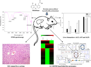
Diclofenac is a hepatotoxic non-steroidal anti-inflammatory drug (NSAID) that affects liver histology and its protein expression levels. Here, we studied the effect of diclofenac on rat liver when co-administrated with either Yersinia enterocolitica strain 8081 serotype O:8 biovar 1B (D*Y) or Lactobacillus fermentum strain 9338 (D*L). Spectroscopic analysis of stool samples showed biotransformation of diclofenac. When compared with each other, D*Y rats lack peaks at 1709 and 1198 cm-1, while D*L rats lack peaks at 1411 cm-1. However, when compared to control, both groups lack peaks at 1379 and 1170 cm-1. Assessment of serum biomarkers of hepatotoxicity indicated significantly altered activities of AST (D*Y: 185.65 ± 8.575 vs Control: 61.9 ± 2.607, D*L: 247.5 ± 5.717 vs Control: 61.9 ± 2.607), ALT (D*Y: 229.8 ± 6.920 vs Control: 70.7 ± 3.109, D*L: 123.75 ± 6.068 vs Control: 70.7 ± 3.109), and ALP (D*Y: 276.4 ± 18.154 vs Control: 320.6 ± 9.829, D*L: 298.5 ± 12.336 vs Control: 320.6 ± 9.829) in IU/L. The analysis of histological alterations showed hepatic sinusoidal dilation with vein congestion and cell infiltration exclusively in D*Y rats along with other histological changes that are common to both test groups, thereby suggesting more pronounced alterations in D*Y rats. Further, LC-MS/MS based label-free quantitation of proteins from liver tissues revealed 74.75% up-regulated, 25.25% down-regulated in D*Y rats and 51.16% up-regulated, 48.84% down-regulated in D*L experiments. The proteomics-identified proteins majorly belonged to metabolism, apoptosis, stress response and redox homeostasis, and detoxification and antioxidant defence that demonstrated the potential damage of rat liver, more pronounced in D*Y rats. Altogether the results are in favor that the administration of lactobacilli somewhat protected the rat hepatic cells against the diclofenac-induced toxicity.
期刊介绍:
Biotechnology Letters is the world’s leading rapid-publication primary journal dedicated to biotechnology as a whole – that is to topics relating to actual or potential applications of biological reactions affected by microbial, plant or animal cells and biocatalysts derived from them.
All relevant aspects of molecular biology, genetics and cell biochemistry, of process and reactor design, of pre- and post-treatment steps, and of manufacturing or service operations are therefore included.
Contributions from industrial and academic laboratories are equally welcome. We also welcome contributions covering biotechnological aspects of regenerative medicine and biomaterials and also cancer biotechnology. Criteria for the acceptance of papers relate to our aim of publishing useful and informative results that will be of value to other workers in related fields.
The emphasis is very much on novelty and immediacy in order to justify rapid publication of authors’ results. It should be noted, however, that we do not normally publish papers (but this is not absolute) that deal with unidentified consortia of microorganisms (e.g. as in activated sludge) as these results may not be easily reproducible in other laboratories.
Papers describing the isolation and identification of microorganisms are not regarded as appropriate but such information can be appended as supporting information to a paper. Papers dealing with simple process development are usually considered to lack sufficient novelty or interest to warrant publication.
 求助内容:
求助内容: 应助结果提醒方式:


