Xenogeneic Collagen Matrix Versus Free Gingival Graft for Augmenting Peri-Implant Keratinized Mucosa Around Dental Implants: A Systematic Review and Meta-Analysis

IF 1.7 Q3 DENTISTRY, ORAL SURGERY & MEDICINE
Momen A. Atieh, Maanas Shah, Abeer Hakam, Suhailah Alshaali, Reem Kasouha, Andrew Tawse-Smith, Nabeel H. M. Alsabeeha
{"title":"Xenogeneic Collagen Matrix Versus Free Gingival Graft for Augmenting Peri-Implant Keratinized Mucosa Around Dental Implants: A Systematic Review and Meta-Analysis","authors":"Momen A. Atieh,&nbsp;Maanas Shah,&nbsp;Abeer Hakam,&nbsp;Suhailah Alshaali,&nbsp;Reem Kasouha,&nbsp;Andrew Tawse-Smith,&nbsp;Nabeel H. M. Alsabeeha","doi":"10.1002/cre2.932","DOIUrl":null,"url":null,"abstract":"<div>\n \n \n <section>\n \n <h3> Objectives</h3>\n \n <p>There is a growing evidence to suggest augmenting peri-implant keratinized mucosa in the presence of ≤ 2 mm of keratinized mucosa. However, the most appropriate surgical technique and augmentation materials have yet to be defined. The aim of this systematic review and meta-analyses was to evaluate the clinical and patient-reported outcomes of augmenting keratinized mucosa around implants using free gingival graft (FGG) versus xenogeneic collagen matrix (XCM) before commencing prosthetic implant treatment.</p>\n </section>\n \n <section>\n \n <h3> Material and Methods</h3>\n \n <p>Electronic databases were searched to identify observational studies comparing implant sites augmented with FGG to those augmented with XCM. The risk of bias was assessed using the Cochrane Collaboration's Risk of Bias tool.</p>\n </section>\n \n <section>\n \n <h3> Results</h3>\n \n <p>Six studies with 174 participants were included in the present review. Of these, 87 participants had FGG, whereas the remaining participants had XCM. At 6 months, sites augmented with FGG were associated with less changes in the gained width of peri-implant keratinized mucosa compared to those augmented with XCM (mean difference 1.06; 95% confidence interval −0.01 to 2.13; <i>p</i> = 0.05). The difference, however, was marginally significant. The difference between the two groups in changes in thickness of peri-implant keratinized mucosa at 6 months was statistically significantly in favor of FGG. On the other hand, XCM had significantly shorter surgical time, lower postoperative pain score, and higher color match compared to FGG.</p>\n </section>\n \n <section>\n \n <h3> Conclusions</h3>\n \n <p>Within the limitation of this review, the augmentation of keratinized mucosa using FGG before the placement of the final prosthesis may have short-term positive effects on soft tissue thickness. XCM might be considered in aesthetically demanding implant sites and where patient comfort or shorter surgical time is a priority. The evidence support, however, is of low to moderate certainty; therefore, further studies are needed to support the findings of the present review.</p>\n </section>\n </div>","PeriodicalId":10203,"journal":{"name":"Clinical and Experimental Dental Research","volume":"10 4","pages":""},"PeriodicalIF":1.7000,"publicationDate":"2024-07-07","publicationTypes":"Journal Article","fieldsOfStudy":null,"isOpenAccess":false,"openAccessPdf":"https://www.ncbi.nlm.nih.gov/pmc/articles/PMC11228352/pdf/","citationCount":"0","resultStr":null,"platform":"Semanticscholar","paperid":null,"PeriodicalName":"Clinical and Experimental Dental Research","FirstCategoryId":"1085","ListUrlMain":"https://onlinelibrary.wiley.com/doi/10.1002/cre2.932","RegionNum":0,"RegionCategory":null,"ArticlePicture":[],"TitleCN":null,"AbstractTextCN":null,"PMCID":null,"EPubDate":"","PubModel":"","JCR":"Q3","JCRName":"DENTISTRY, ORAL SURGERY & MEDICINE","Score":null,"Total":0}
引用次数: 0

Abstract

Objectives

There is a growing evidence to suggest augmenting peri-implant keratinized mucosa in the presence of ≤ 2 mm of keratinized mucosa. However, the most appropriate surgical technique and augmentation materials have yet to be defined. The aim of this systematic review and meta-analyses was to evaluate the clinical and patient-reported outcomes of augmenting keratinized mucosa around implants using free gingival graft (FGG) versus xenogeneic collagen matrix (XCM) before commencing prosthetic implant treatment.

Material and Methods

Electronic databases were searched to identify observational studies comparing implant sites augmented with FGG to those augmented with XCM. The risk of bias was assessed using the Cochrane Collaboration's Risk of Bias tool.

Results

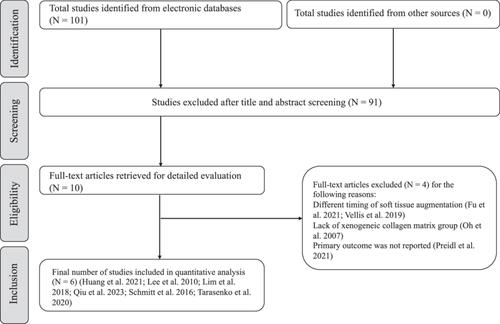
Six studies with 174 participants were included in the present review. Of these, 87 participants had FGG, whereas the remaining participants had XCM. At 6 months, sites augmented with FGG were associated with less changes in the gained width of peri-implant keratinized mucosa compared to those augmented with XCM (mean difference 1.06; 95% confidence interval −0.01 to 2.13; p = 0.05). The difference, however, was marginally significant. The difference between the two groups in changes in thickness of peri-implant keratinized mucosa at 6 months was statistically significantly in favor of FGG. On the other hand, XCM had significantly shorter surgical time, lower postoperative pain score, and higher color match compared to FGG.

Conclusions

Within the limitation of this review, the augmentation of keratinized mucosa using FGG before the placement of the final prosthesis may have short-term positive effects on soft tissue thickness. XCM might be considered in aesthetically demanding implant sites and where patient comfort or shorter surgical time is a priority. The evidence support, however, is of low to moderate certainty; therefore, further studies are needed to support the findings of the present review.
Abstract Image

异种胶原基质与游离牙龈移植用于牙种植体周围角质化粘膜的增量:系统回顾与元分析》。
目的:越来越多的证据表明,如果种植体周围的角化粘膜少于 2 毫米,则建议增量种植体周围的角化粘膜。然而,最合适的手术技术和增量材料仍有待确定。本系统综述和荟萃分析的目的是评估在开始修复种植体治疗前使用游离龈移植(FGG)与异种胶原基质(XCM)增量种植体周围角化粘膜的临床和患者报告结果:对电子数据库进行检索,以确定对使用 FGG 增量的种植部位与使用 XCM 增量的种植部位进行比较的观察性研究。使用 Cochrane 协作组织的偏倚风险工具对偏倚风险进行了评估:本综述纳入了六项研究,共有 174 名参与者。其中,87 名参与者接受了 FGG 治疗,其余参与者接受了 XCM 治疗。6 个月后,与使用 XCM 增量的部位相比,使用 FGG 增量的部位种植体周围角化粘膜增宽的变化较小(平均差异为 1.06;95% 置信区间为-0.01 至 2.13;P = 0.05)。不过,差异略微显著。在 6 个月时,两组种植体周围角化粘膜厚度变化的差异在统计学上明显有利于 FGG。另一方面,与 FGG 相比,XCM 的手术时间明显更短,术后疼痛评分更低,颜色匹配度更高:在这篇综述的局限性范围内,在植入最终假体前使用 FGG 增加角化粘膜可能会对软组织厚度产生短期的积极影响。在对美观要求较高的种植部位,以及患者舒适度或手术时间较短的情况下,可以考虑使用 XCM。不过,证据支持的确定性为中低,因此需要进一步的研究来支持本综述的结论。
本文章由计算机程序翻译,如有差异,请以英文原文为准。
求助全文
约1分钟内获得全文 求助全文
来源期刊
Clinical and Experimental Dental Research
Clinical and Experimental Dental Research DENTISTRY, ORAL SURGERY & MEDICINE-
CiteScore
3.30
自引率
5.60%
发文量
165
审稿时长
26 weeks
期刊介绍: Clinical and Experimental Dental Research aims to provide open access peer-reviewed publications of high scientific quality representing original clinical, diagnostic or experimental work within all disciplines and fields of oral medicine and dentistry. The scope of Clinical and Experimental Dental Research comprises original research material on the anatomy, physiology and pathology of oro-facial, oro-pharyngeal and maxillofacial tissues, and functions and dysfunctions within the stomatognathic system, and the epidemiology, aetiology, prevention, diagnosis, prognosis and therapy of diseases and conditions that have an effect on the homeostasis of the mouth, jaws, and closely associated structures, as well as the healing and regeneration and the clinical aspects of replacement of hard and soft tissues with biomaterials, and the rehabilitation of stomatognathic functions. Studies that bring new knowledge on how to advance health on the individual or public health levels, including interactions between oral and general health and ill-health are welcome.
×
引用
GB/T 7714-2015
复制
MLA
复制
APA
复制
导出至
BibTeX EndNote RefMan NoteFirst NoteExpress
×
提示
您的信息不完整,为了账户安全,请先补充。
现在去补充
×
提示
您因"违规操作"
具体请查看互助需知
我知道了
×
提示
确定
请完成安全验证×
copy
已复制链接
快去分享给好友吧!
我知道了
右上角分享
点击右上角分享
0
联系我们:info@booksci.cn Book学术提供免费学术资源搜索服务,方便国内外学者检索中英文文献。致力于提供最便捷和优质的服务体验。 Copyright © 2023 布克学术 All rights reserved.
京ICP备2023020795号-1
ghs 京公网安备 11010802042870号
Book学术文献互助
Book学术文献互助群
群 号:481959085
Book学术官方微信