Morphometric Analysis of Serotoninergic Structures in the Nervous System of the Planarian Schmidtea mediterranea
Abstract
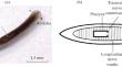
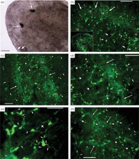
The planarian nervous system is represented by a cephalic ganglion in the anterior part of the body and a pair of well-defined ventral nerve trunks extending along the entire body of the animal. The serotoninergic components of the nervous system are determined by the indirect immunocytochemical staining of the total preparations of the planarian Schmidtea mediterranea tissues, followed by analysis by fluorescent microscopy. The presence of serotoninergic components is found in the central and peripheral parts of the nervous system of the planarian S. mediterranea. The morphological parameters of serotonin-immunopositive structures are measured and the number of neurons in the cerebral ganglion is counted. The measurements are carried out on micrographs from stained total preparations obtained using a digital camera. The size of serotonin neurons in three areas of the body, the thickness of nerve trunks and cerebral ganglion, and the distance between nerve trunks and commissures are considered. For the first time, new quantitative data characterizing the morphological properties of the nervous system of the planarian S. mediterranea have been obtained. The regeneration of planarian eyes after decapitation and serotonin exposure is also studied. It is found that exogenous serotonin at a concentration of 0.01–1 μM accelerated eye differentiation during regeneration of the head end of S. mediterranea.
 求助内容:
求助内容: 应助结果提醒方式:


