Suppression of Aseptic Inflammation Reduces the Severity of Pulmonary Artery Remodeling and Improves the Clinical Course of Experimental Chronic Thromboembolic Pulmonary Hypertension
A. A. Karpov, A. A. Krylov, L. A. Shilenko, A. M. Mihailova, D. D. Vaulina, D. Yu. Ivkin, N. P. Isakova, A. V. Vorotilov, N. Yu. Semenova, V. A. Zinserling, M. M. Galagudza
{"title":"Suppression of Aseptic Inflammation Reduces the Severity of Pulmonary Artery Remodeling and Improves the Clinical Course of Experimental Chronic Thromboembolic Pulmonary Hypertension","authors":"A. A. Karpov, A. A. Krylov, L. A. Shilenko, A. M. Mihailova, D. D. Vaulina, D. Yu. Ivkin, N. P. Isakova, A. V. Vorotilov, N. Yu. Semenova, V. A. Zinserling, M. M. Galagudza","doi":"10.1134/s002209302403030x","DOIUrl":null,"url":null,"abstract":"<h3 data-test=\"abstract-sub-heading\">Abstract</h3><p>Chronic thromboembolic pulmonary hypertension (CTEPH) is a\ncomplication of pulmonary embolism, characterized by high blood\npressure in the pulmonary artery combined with impaired lysis of fibrin\nclots. Previously, the presence of aseptic inflammation in CTEPH\nwas found in the wall of the pulmonary artery branches, as well\nas perivascularly. However, the role of this inflammation in shaping CTEPH\nis unknown. The aim of the work was to study the effect of aseptic\ninflammation on CTEPH formation and progression. The experiments\nwere carried out on male Wistar rats (<i>n</i> =\n54). The CTEPH model was reproduced by repeated intravenous administration\nof partially biodegradable microspheres (MS). Immediately after\nthe last MS administration, all animals were randomly allocated\ninto four groups: (1) control (c.CTEPH) group—physiological saline\nwas administered intramuscularly (i.m.) for 6 weeks; (2) low-dose\nprednisolone (LDP) group—prednisolone was administered i.m. at a\ndose of 1.5 mg/kg; (3) high-dose prednisolone (HDP) group—prednisolone\nwas administered i.m. at a dose of 6 mg/kg; (4) healthy or intact\n(INT) group. After 6 weeks, there were performed the treadmill test, transthoracic\nechocardiography, cardiac catheterization with blood pressure manometry,\nand lung tissue histological examination. In a separate series of\nexperiments, the degree of vascular wall and perivascular space\ninflammatory infiltration was assessed immunohistochemically. In\nLDP group, the vascular wall hypertrophy index (HI) and the percentage\nof collagen fibers in the vascular wall were reduced compared to\nthe control group, with the HI being reduced significantly greater\nthan in HDP group. In the latter, there was revealed a positive\neffect of high-dose prednisolone on the percentage of collagen fibers\nin the vascular wall, with this parameter being non-significantly\ndifferent from that in intact animals. By immunohistochemical data,\nlow-dose prednisolone effectively suppressed inflammatory infiltration\nof the vascular wall and perivascular space. Thus, we revealed the\nability of low-dose prednisolone to reduce the degree of remodeling\nof the pulmonary artery branches by suppressing aseptic inflammation.</p>","PeriodicalId":15805,"journal":{"name":"Journal of Evolutionary Biochemistry and Physiology","volume":"25 1","pages":""},"PeriodicalIF":0.5000,"publicationDate":"2024-06-26","publicationTypes":"Journal Article","fieldsOfStudy":null,"isOpenAccess":false,"openAccessPdf":"","citationCount":"0","resultStr":null,"platform":"Semanticscholar","paperid":null,"PeriodicalName":"Journal of Evolutionary Biochemistry and Physiology","FirstCategoryId":"99","ListUrlMain":"https://doi.org/10.1134/s002209302403030x","RegionNum":4,"RegionCategory":"生物学","ArticlePicture":[],"TitleCN":null,"AbstractTextCN":null,"PMCID":null,"EPubDate":"","PubModel":"","JCR":"Q4","JCRName":"BIOCHEMISTRY & MOLECULAR BIOLOGY","Score":null,"Total":0}
引用次数: 0
Abstract
Chronic thromboembolic pulmonary hypertension (CTEPH) is a
complication of pulmonary embolism, characterized by high blood
pressure in the pulmonary artery combined with impaired lysis of fibrin
clots. Previously, the presence of aseptic inflammation in CTEPH
was found in the wall of the pulmonary artery branches, as well
as perivascularly. However, the role of this inflammation in shaping CTEPH
is unknown. The aim of the work was to study the effect of aseptic
inflammation on CTEPH formation and progression. The experiments
were carried out on male Wistar rats (n =
54). The CTEPH model was reproduced by repeated intravenous administration
of partially biodegradable microspheres (MS). Immediately after
the last MS administration, all animals were randomly allocated
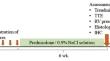
into four groups: (1) control (c.CTEPH) group—physiological saline
was administered intramuscularly (i.m.) for 6 weeks; (2) low-dose
prednisolone (LDP) group—prednisolone was administered i.m. at a
dose of 1.5 mg/kg; (3) high-dose prednisolone (HDP) group—prednisolone
was administered i.m. at a dose of 6 mg/kg; (4) healthy or intact
(INT) group. After 6 weeks, there were performed the treadmill test, transthoracic
echocardiography, cardiac catheterization with blood pressure manometry,
and lung tissue histological examination. In a separate series of
experiments, the degree of vascular wall and perivascular space
inflammatory infiltration was assessed immunohistochemically. In
LDP group, the vascular wall hypertrophy index (HI) and the percentage
of collagen fibers in the vascular wall were reduced compared to
the control group, with the HI being reduced significantly greater
than in HDP group. In the latter, there was revealed a positive
effect of high-dose prednisolone on the percentage of collagen fibers
in the vascular wall, with this parameter being non-significantly
different from that in intact animals. By immunohistochemical data,
low-dose prednisolone effectively suppressed inflammatory infiltration
of the vascular wall and perivascular space. Thus, we revealed the
ability of low-dose prednisolone to reduce the degree of remodeling
of the pulmonary artery branches by suppressing aseptic inflammation.
期刊介绍:
Journal of Evolutionary Biochemistry and Physiology publishes original experimental and theoretical and review articles related to evolution of the main forms of metabolism in connection with life origin; comparative and ontogenetic physiology and biochemistry, biochemical evolution of animal world; as well as evolution of functions; morphology, pharmacology, pathophysiology and ecological physiology. The journal welcomes manuscripts from all countries in the English or Russian language.
 求助内容:
求助内容: 应助结果提醒方式:


