Sricharan S Veeturi, Arshaq Saleem, Diego J Ojeda, Elena Sagues, Sebastian Sanchez, Andres Gudino, Elad I Levy, David Hasan, Adnan H Siddiqui, Vincent M Tutino, Edgar A Samaniego
{"title":"Radiomics-Based Predictive Nomogram for Assessing the Risk of Intracranial Aneurysms.","authors":"Sricharan S Veeturi, Arshaq Saleem, Diego J Ojeda, Elena Sagues, Sebastian Sanchez, Andres Gudino, Elad I Levy, David Hasan, Adnan H Siddiqui, Vincent M Tutino, Edgar A Samaniego","doi":"10.1007/s12975-024-01268-3","DOIUrl":null,"url":null,"abstract":"<p><p>Aneurysm wall enhancement (AWE) has the potential to be used as an imaging biomarker for the risk stratification of intracranial aneurysms (IAs). Radiomics provides a refined approach to quantify and further characterize AWE's textural features. This study examines the performance of AWE quantification combined with clinical information in detecting symptomatic IAs. Ninety patients harboring 104 IAs (29 symptomatic and 75 asymptomatic) underwent high-resolution magnetic resonance imaging (HR-MRI). The assessment of AWE was performed using two different methods: 3D-AWE mapping and composite radiomics-based score (RadScore). The dataset was split into training and testing subsets. The testing set was used to build two different nomograms using each modality of AWE assessment combined with patients' clinical information and aneurysm morphological data. Finally, each nomogram was evaluated on an independent testing set. A total of 22 radiomic features were significantly different between symptomatic and asymptomatic IAs. The 3D-AWE mapping nomogram achieved an area under the curve (AUC) of 0.77 (63% accuracy, 78% sensitivity, and 58% specificity). The RadScore nomogram exhibited a better performance, achieving an AUC of 0.83 (77% accuracy, 89% sensitivity, and 73% specificity). The comprehensive analysis of IAs with the quantification of AWE data through radiomic analysis, patient clinical information, and morphological aneurysm metrics achieves a high accuracy in detecting symptomatic IA status.</p>","PeriodicalId":23237,"journal":{"name":"Translational Stroke Research","volume":" ","pages":"79-87"},"PeriodicalIF":3.8000,"publicationDate":"2025-02-01","publicationTypes":"Journal Article","fieldsOfStudy":null,"isOpenAccess":false,"openAccessPdf":"","citationCount":"0","resultStr":null,"platform":"Semanticscholar","paperid":null,"PeriodicalName":"Translational Stroke Research","FirstCategoryId":"3","ListUrlMain":"https://doi.org/10.1007/s12975-024-01268-3","RegionNum":2,"RegionCategory":"医学","ArticlePicture":[],"TitleCN":null,"AbstractTextCN":null,"PMCID":null,"EPubDate":"2024/7/2 0:00:00","PubModel":"Epub","JCR":"Q1","JCRName":"CLINICAL NEUROLOGY","Score":null,"Total":0}
引用次数: 0
Abstract
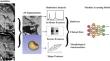
Aneurysm wall enhancement (AWE) has the potential to be used as an imaging biomarker for the risk stratification of intracranial aneurysms (IAs). Radiomics provides a refined approach to quantify and further characterize AWE's textural features. This study examines the performance of AWE quantification combined with clinical information in detecting symptomatic IAs. Ninety patients harboring 104 IAs (29 symptomatic and 75 asymptomatic) underwent high-resolution magnetic resonance imaging (HR-MRI). The assessment of AWE was performed using two different methods: 3D-AWE mapping and composite radiomics-based score (RadScore). The dataset was split into training and testing subsets. The testing set was used to build two different nomograms using each modality of AWE assessment combined with patients' clinical information and aneurysm morphological data. Finally, each nomogram was evaluated on an independent testing set. A total of 22 radiomic features were significantly different between symptomatic and asymptomatic IAs. The 3D-AWE mapping nomogram achieved an area under the curve (AUC) of 0.77 (63% accuracy, 78% sensitivity, and 58% specificity). The RadScore nomogram exhibited a better performance, achieving an AUC of 0.83 (77% accuracy, 89% sensitivity, and 73% specificity). The comprehensive analysis of IAs with the quantification of AWE data through radiomic analysis, patient clinical information, and morphological aneurysm metrics achieves a high accuracy in detecting symptomatic IA status.
期刊介绍:
Translational Stroke Research covers basic, translational, and clinical studies. The Journal emphasizes novel approaches to help both to understand clinical phenomenon through basic science tools, and to translate basic science discoveries into the development of new strategies for the prevention, assessment, treatment, and enhancement of central nervous system repair after stroke and other forms of neurotrauma.
Translational Stroke Research focuses on translational research and is relevant to both basic scientists and physicians, including but not restricted to neuroscientists, vascular biologists, neurologists, neuroimagers, and neurosurgeons.
 求助内容:
求助内容: 应助结果提醒方式:


