Uncorrected and subcutaneous fat-corrected echo intensities are similarly associated with magnetic resonance imaging per cent fat

Benjamin Rush, Sujay Garlapati, Jevin Lortie, Katie Osterbauer, Timothy J. Colgan, Daiki Tamada, Toby C. Campbell, Anne Traynor, Ticiana Leal, Kenneth Lee, Scott B. Reeder, Adam J. Kuchnia
{"title":"Uncorrected and subcutaneous fat-corrected echo intensities are similarly associated with magnetic resonance imaging per cent fat","authors":"Benjamin Rush,&nbsp;Sujay Garlapati,&nbsp;Jevin Lortie,&nbsp;Katie Osterbauer,&nbsp;Timothy J. Colgan,&nbsp;Daiki Tamada,&nbsp;Toby C. Campbell,&nbsp;Anne Traynor,&nbsp;Ticiana Leal,&nbsp;Kenneth Lee,&nbsp;Scott B. Reeder,&nbsp;Adam J. Kuchnia","doi":"10.1002/rco2.92","DOIUrl":null,"url":null,"abstract":"<div>\n \n \n <section>\n \n <h3> Background</h3>\n \n <p>Establishing interchangeable biomedical imaging-based measures to assess myosteatosis clinically may lead to the prevention of muscle wasting, yet neither a consensus measure nor a conversion between measures exists. Ultrasound echo intensity (EI) potentially assesses myosteatosis, but subcutaneous adipose tissue (SAT) thickness and user force application have been shown to influence EI. Although correction factors exist to adjust EI for SAT thickness, they are modelled against poor or no reference measures. Modelling EI corrections against a robust reference measure of myosteatosis, like magnetic resonance imaging (MRI)-based proton density fat fraction (PDFF), is necessary for EI's clinical application.</p>\n </section>\n \n <section>\n \n <h3> Methods</h3>\n \n <p>Healthy young adults, healthy older adults, and older adults undergoing treatment for lung cancer (<i>n</i> = 10 per group with 50% females) had PDFF and EI at 0, 5, 10, and 15 N measured on their right rectus femoris (RF). We compared EI, SAT thickness, and RF thickness between forces and groups and assessed the relationships between EI adjusted by four different correction factors and PDFF.</p>\n </section>\n \n <section>\n \n <h3> Results</h3>\n \n <p>The mean age of our sample was 48.63 ± 19.68 years and had a body mass index of 25.21 ± 5.19 kg/m<sup>2</sup>. The correlation between PDFF and raw EI was <i>r</i> = 0.59 (<i>P</i> &lt; 0.001) with negligible increases by previously published correction factors (Young: 0.62, <i>P</i> &lt; 0.001; Neto Müller: 0.61, <i>P</i> &lt; 0.001). EI, SAT thickness, and RF thickness did not significantly differ between forces (<i>χ</i><sup>2</sup> = 0.31, <i>P</i> = 0.957; <i>χ</i><sup>2</sup> = 2.39, <i>P</i> = 0.496; and <i>χ</i><sup>2</sup> = 7.75, <i>P</i> = 0.051, respectively). EI and PDFF were significantly lower among young healthy adults compared with older adult groups (<i>χ</i><sup>2</sup> = 12.88, <i>P</i> = 0.002, and <i>χ</i><sup>2</sup> = 9.13, <i>P</i> = 0.010, respectively).</p>\n </section>\n \n <section>\n \n <h3> Conclusions</h3>\n \n <p>EI is correlated with PDFF regardless of force with no improvement from previously published correction factors. Our results suggest that EI is clinically useful and influenced by fat content, yet correction factors must account for more than SAT thickness alone and require further investigation.</p>\n </section>\n </div>","PeriodicalId":73544,"journal":{"name":"JCSM rapid communications","volume":null,"pages":null},"PeriodicalIF":0.0000,"publicationDate":"2024-05-07","publicationTypes":"Journal Article","fieldsOfStudy":null,"isOpenAccess":false,"openAccessPdf":"https://onlinelibrary.wiley.com/doi/epdf/10.1002/rco2.92","citationCount":"0","resultStr":null,"platform":"Semanticscholar","paperid":null,"PeriodicalName":"JCSM rapid communications","FirstCategoryId":"1085","ListUrlMain":"https://onlinelibrary.wiley.com/doi/10.1002/rco2.92","RegionNum":0,"RegionCategory":null,"ArticlePicture":[],"TitleCN":null,"AbstractTextCN":null,"PMCID":null,"EPubDate":"","PubModel":"","JCR":"","JCRName":"","Score":null,"Total":0}
引用次数: 0

Abstract

Background

Establishing interchangeable biomedical imaging-based measures to assess myosteatosis clinically may lead to the prevention of muscle wasting, yet neither a consensus measure nor a conversion between measures exists. Ultrasound echo intensity (EI) potentially assesses myosteatosis, but subcutaneous adipose tissue (SAT) thickness and user force application have been shown to influence EI. Although correction factors exist to adjust EI for SAT thickness, they are modelled against poor or no reference measures. Modelling EI corrections against a robust reference measure of myosteatosis, like magnetic resonance imaging (MRI)-based proton density fat fraction (PDFF), is necessary for EI's clinical application.

Methods

Healthy young adults, healthy older adults, and older adults undergoing treatment for lung cancer (n = 10 per group with 50% females) had PDFF and EI at 0, 5, 10, and 15 N measured on their right rectus femoris (RF). We compared EI, SAT thickness, and RF thickness between forces and groups and assessed the relationships between EI adjusted by four different correction factors and PDFF.

Results

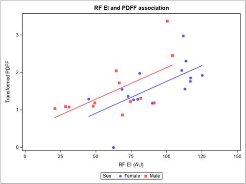
The mean age of our sample was 48.63 ± 19.68 years and had a body mass index of 25.21 ± 5.19 kg/m2. The correlation between PDFF and raw EI was r = 0.59 (P < 0.001) with negligible increases by previously published correction factors (Young: 0.62, P < 0.001; Neto Müller: 0.61, P < 0.001). EI, SAT thickness, and RF thickness did not significantly differ between forces (χ2 = 0.31, P = 0.957; χ2 = 2.39, P = 0.496; and χ2 = 7.75, P = 0.051, respectively). EI and PDFF were significantly lower among young healthy adults compared with older adult groups (χ2 = 12.88, P = 0.002, and χ2 = 9.13, P = 0.010, respectively).

Conclusions

EI is correlated with PDFF regardless of force with no improvement from previously published correction factors. Our results suggest that EI is clinically useful and influenced by fat content, yet correction factors must account for more than SAT thickness alone and require further investigation.

Abstract Image

未校正回波强度和皮下脂肪校正回波强度与磁共振成像脂肪百分比的关系相似
背景 建立可互换的基于生物医学成像的临床评估方法来评估肌肉骨质疏松症,可能有助于预防肌肉萎缩,但目前既没有达成共识的方法,也不存在不同方法之间的转换。超声回波强度(EI)可评估肌骨软化症,但已证明皮下脂肪组织(SAT)厚度和使用者施力会影响 EI。尽管存在根据 SAT 厚度调整 EI 的校正因子,但它们都是根据较差的或没有参考测量值的情况建模的。根据可靠的肌骨质疏松症参考指标(如基于磁共振成像(MRI)的质子密度脂肪分数(PDFF))来建立 EI 修正模型,对于 EI 的临床应用非常必要。 方法 对健康的年轻人、健康的老年人和正在接受肺癌治疗的老年人(每组 10 人,女性占 50%)的右股直肌(RF)进行 0、5、10 和 15 N 的质子密度脂肪分数(PDFF)和 EI 测量。我们比较了不同力量和组间的 EI、SAT 厚度和 RF 厚度,并评估了经四种不同校正因子调整的 EI 与 PDFF 之间的关系。 结果 样本的平均年龄为 48.63 ± 19.68 岁,体重指数为 25.21 ± 5.19 kg/m2。PDFF 与原始 EI 之间的相关性为 r = 0.59(P < 0.001),与之前公布的校正因子(Young:0.62,P < 0.001;Neto Müller:0.61,P < 0.001)的相关性几乎可以忽略不计。不同力量之间的 EI、SAT 厚度和 RF 厚度差异不大(分别为 χ2 = 0.31,P = 0.957;χ2 = 2.39,P = 0.496;χ2 = 7.75,P = 0.051)。与老年人组相比,年轻健康成人的 EI 和 PDFF 明显较低(分别为 χ2 = 12.88,P = 0.002 和 χ2 = 9.13,P = 0.010)。 结论 无论力量大小,EI 都与 PDFF 相关,与之前公布的校正因子相比没有改进。我们的结果表明,EI 在临床上是有用的,并受脂肪含量的影响,但校正因子必须比 SAT 厚度单独考虑更多因素,因此需要进一步研究。
本文章由计算机程序翻译,如有差异,请以英文原文为准。
求助全文
约1分钟内获得全文 求助全文
来源期刊
自引率
0.00%
发文量
0
审稿时长
10 weeks
文献相关原料
公司名称 产品信息 采购帮参考价格
×
引用
GB/T 7714-2015
复制
MLA
复制
APA
复制
导出至
BibTeX EndNote RefMan NoteFirst NoteExpress
×
提示
您的信息不完整,为了账户安全,请先补充。
现在去补充
×
提示
您因"违规操作"
具体请查看互助需知
我知道了
×
提示
确定
请完成安全验证×
copy
已复制链接
快去分享给好友吧!
我知道了
右上角分享
点击右上角分享
0
联系我们:info@booksci.cn Book学术提供免费学术资源搜索服务,方便国内外学者检索中英文文献。致力于提供最便捷和优质的服务体验。 Copyright © 2023 布克学术 All rights reserved.
京ICP备2023020795号-1
ghs 京公网安备 11010802042870号
Book学术文献互助
Book学术文献互助群
群 号:481959085
Book学术官方微信