Comparison of the effectiveness of contrast-enhanced mammography in detecting malignant lesions in patients with extremely dense breasts compared to the all-densities population.
Anna Grażyńska, Agnieszka Niewiadomska, Aleksander J Owczarek, Mateusz Winder, Jakub Hołda, Olga Zwolińska, Anna Barczyk-Gutkowska, Sandra Modlińska, Andrzej Lorek, Aleksandra Kuźbińska, Katarzyna Steinhof-Radwańska
{"title":"Comparison of the effectiveness of contrast-enhanced mammography in detecting malignant lesions in patients with extremely dense breasts compared to the all-densities population.","authors":"Anna Grażyńska, Agnieszka Niewiadomska, Aleksander J Owczarek, Mateusz Winder, Jakub Hołda, Olga Zwolińska, Anna Barczyk-Gutkowska, Sandra Modlińska, Andrzej Lorek, Aleksandra Kuźbińska, Katarzyna Steinhof-Radwańska","doi":"10.5114/pjr/186180","DOIUrl":null,"url":null,"abstract":"<p><strong>Purpose: </strong>To assess the effectiveness of contrast-enhanced mammography (CEM) recombinant images in detecting malignant lesions in patients with extremely dense breasts compared to the all-densities population.</p><p><strong>Material and methods: </strong>792 patients with 808 breast lesions, in whom the final decision on core-needle biopsy was made based on CEM, and who received the result of histopathological examination, were qualified for a single-centre, retrospective study. Patient electronic records and imaging examinations were reviewed to establish demographics, clinical and imaging findings, and histopathology results. The CEM images were reassessed and assigned to the appropriate American College of Radiology (ACR) density categories.</p><p><strong>Results: </strong>Extremely dense breasts were present in 86 (10.9%) patients. Histopathological examination confirmed the presence of malignant lesions in 52.6% of cases in the entire group of patients and 43% in the group of extremely dense breasts. CEM incorrectly classified the lesion as false negative in 16/425 (3.8%) cases for the whole group, and in 1/37 (2.7%) cases for extremely dense breasts. The sensitivity of CEM for the group of all patients was 96.2%, specificity - 60%, positive predictive values (PPV) - 72.8%, and negative predictive values (NPV) - 93.5%. In the group of patients with extremely dense breasts, the sensitivity of the method was 97.3%, specificity - 59.2%, PPV - 64.3%, and NPV - 96.7%.</p><p><strong>Conclusions: </strong>CEM is characterised by high sensitivity and NPV in detecting malignant lesions regardless of the type of breast density. In patients with extremely dense breasts, CEM could serve as a complementary or additional examination in the absence or low availability of MRI.</p>","PeriodicalId":94174,"journal":{"name":"Polish journal of radiology","volume":"89 ","pages":"e240-e248"},"PeriodicalIF":0.0000,"publicationDate":"2024-05-15","publicationTypes":"Journal Article","fieldsOfStudy":null,"isOpenAccess":false,"openAccessPdf":"https://www.ncbi.nlm.nih.gov/pmc/articles/PMC11210381/pdf/","citationCount":"0","resultStr":null,"platform":"Semanticscholar","paperid":null,"PeriodicalName":"Polish journal of radiology","FirstCategoryId":"1085","ListUrlMain":"https://doi.org/10.5114/pjr/186180","RegionNum":0,"RegionCategory":null,"ArticlePicture":[],"TitleCN":null,"AbstractTextCN":null,"PMCID":null,"EPubDate":"2024/1/1 0:00:00","PubModel":"eCollection","JCR":"","JCRName":"","Score":null,"Total":0}
引用次数: 0
Abstract
Purpose: To assess the effectiveness of contrast-enhanced mammography (CEM) recombinant images in detecting malignant lesions in patients with extremely dense breasts compared to the all-densities population.
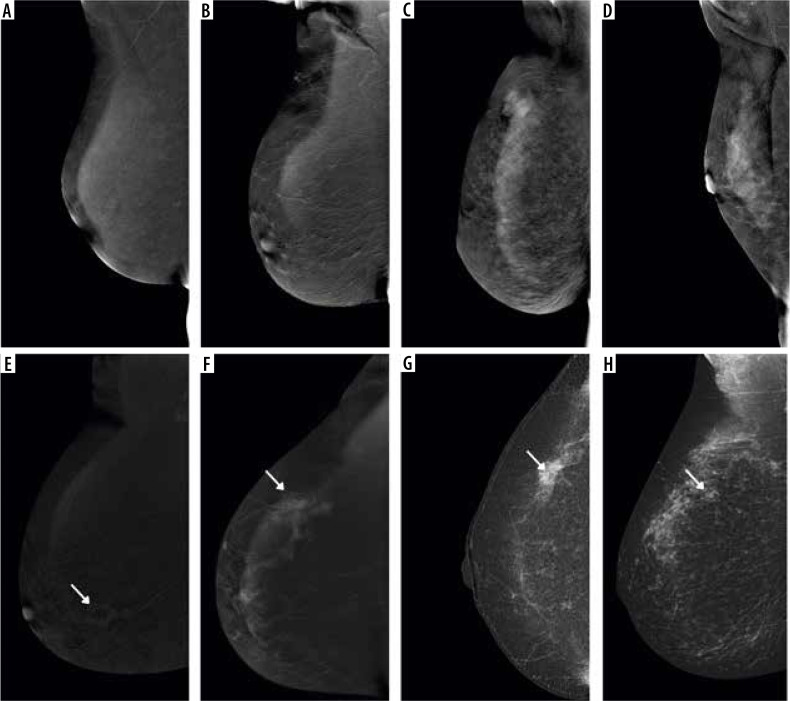
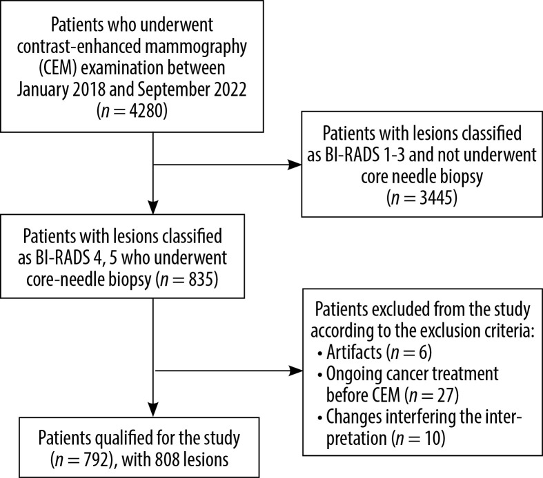
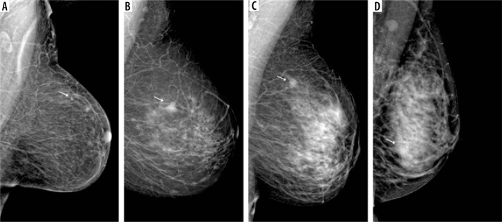
Material and methods: 792 patients with 808 breast lesions, in whom the final decision on core-needle biopsy was made based on CEM, and who received the result of histopathological examination, were qualified for a single-centre, retrospective study. Patient electronic records and imaging examinations were reviewed to establish demographics, clinical and imaging findings, and histopathology results. The CEM images were reassessed and assigned to the appropriate American College of Radiology (ACR) density categories.
Results: Extremely dense breasts were present in 86 (10.9%) patients. Histopathological examination confirmed the presence of malignant lesions in 52.6% of cases in the entire group of patients and 43% in the group of extremely dense breasts. CEM incorrectly classified the lesion as false negative in 16/425 (3.8%) cases for the whole group, and in 1/37 (2.7%) cases for extremely dense breasts. The sensitivity of CEM for the group of all patients was 96.2%, specificity - 60%, positive predictive values (PPV) - 72.8%, and negative predictive values (NPV) - 93.5%. In the group of patients with extremely dense breasts, the sensitivity of the method was 97.3%, specificity - 59.2%, PPV - 64.3%, and NPV - 96.7%.
Conclusions: CEM is characterised by high sensitivity and NPV in detecting malignant lesions regardless of the type of breast density. In patients with extremely dense breasts, CEM could serve as a complementary or additional examination in the absence or low availability of MRI.

 求助内容:
求助内容: 应助结果提醒方式:


