Marco Isaac, Dina Mohamed ElBeshlawy, Ahmed Elsobki, Dina Fahim Ahmed, Sarah Mohammed Kenawy
{"title":"Correlation between cone-beam computed tomographic findings and the apnea-hypopnea index in obstructive sleep apnea patients: A cross-sectional study.","authors":"Marco Isaac, Dina Mohamed ElBeshlawy, Ahmed Elsobki, Dina Fahim Ahmed, Sarah Mohammed Kenawy","doi":"10.5624/isd.20230249","DOIUrl":null,"url":null,"abstract":"<p><strong>Purpose: </strong>The aim of this study was to explore the correlations of cone-beam computed tomographic findings with the apnea-hypopnea index in patients with obstructive sleep apnea.</p><p><strong>Materials and methods: </strong>Forty patients with obstructive sleep apnea were selected from the ear-nose-throat (ENT) outpatient clinic, Faculty of Medicine, Mansoura University. Cone-beam computed tomography was performed for each patient at the end of both inspiration and expiration. Polysomnography was carried out, and the apnea-hypopnea index was obtained. Linear measurements, including cross-sectional area and the SNA and SNB angles, were obtained. Four oral and maxillofacial radiologists categorized pharyngeal and retropalatal airway morphology and calculated the airway length and volume. Continuous data were tested for normality using the Kolmogorov-Smirnov test and reported as the mean and standard deviation or as the median and range. Categorical data were presented as numbers and percentages, and the significance level was set at <i>P</i><0.05.</p><p><strong>Results: </strong>The minimal value of the cross-sectional area, SNB angle, and airway morphology at the end of inspiration demonstrated a statistically significant association (<i>P</i><0.05) with the apnea-hypopnea index, with excellent agreement. No statistically significant difference was found in the airway volume, other linear measurements, or retropalatal airway morphology.</p><p><strong>Conclusion: </strong>Cone-beam computed tomographic measurements in obstructive sleep apnea patients may be used as a supplement to a novel radiographic classification corresponding to the established clinical apnea-hypopnea index classification.</p>","PeriodicalId":51714,"journal":{"name":"Imaging Science in Dentistry","volume":"54 2","pages":"147-157"},"PeriodicalIF":2.1000,"publicationDate":"2024-06-01","publicationTypes":"Journal Article","fieldsOfStudy":null,"isOpenAccess":false,"openAccessPdf":"https://www.ncbi.nlm.nih.gov/pmc/articles/PMC11211029/pdf/","citationCount":"0","resultStr":null,"platform":"Semanticscholar","paperid":null,"PeriodicalName":"Imaging Science in Dentistry","FirstCategoryId":"1085","ListUrlMain":"https://doi.org/10.5624/isd.20230249","RegionNum":0,"RegionCategory":null,"ArticlePicture":[],"TitleCN":null,"AbstractTextCN":null,"PMCID":null,"EPubDate":"2024/4/2 0:00:00","PubModel":"Epub","JCR":"Q3","JCRName":"DENTISTRY, ORAL SURGERY & MEDICINE","Score":null,"Total":0}
引用次数: 0
Abstract
Purpose: The aim of this study was to explore the correlations of cone-beam computed tomographic findings with the apnea-hypopnea index in patients with obstructive sleep apnea.
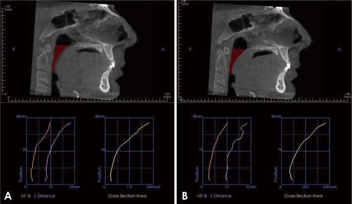
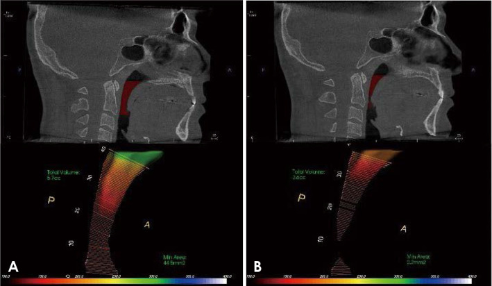
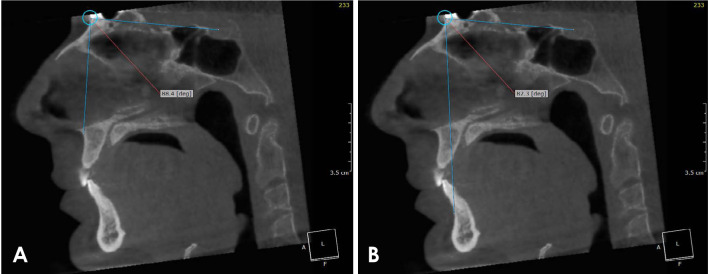
Materials and methods: Forty patients with obstructive sleep apnea were selected from the ear-nose-throat (ENT) outpatient clinic, Faculty of Medicine, Mansoura University. Cone-beam computed tomography was performed for each patient at the end of both inspiration and expiration. Polysomnography was carried out, and the apnea-hypopnea index was obtained. Linear measurements, including cross-sectional area and the SNA and SNB angles, were obtained. Four oral and maxillofacial radiologists categorized pharyngeal and retropalatal airway morphology and calculated the airway length and volume. Continuous data were tested for normality using the Kolmogorov-Smirnov test and reported as the mean and standard deviation or as the median and range. Categorical data were presented as numbers and percentages, and the significance level was set at P<0.05.
Results: The minimal value of the cross-sectional area, SNB angle, and airway morphology at the end of inspiration demonstrated a statistically significant association (P<0.05) with the apnea-hypopnea index, with excellent agreement. No statistically significant difference was found in the airway volume, other linear measurements, or retropalatal airway morphology.
Conclusion: Cone-beam computed tomographic measurements in obstructive sleep apnea patients may be used as a supplement to a novel radiographic classification corresponding to the established clinical apnea-hypopnea index classification.

 求助内容:
求助内容: 应助结果提醒方式:


