Natalia P. Silva , Charles S. da Costa , Kayke L. Barbosa , Cidália de F. Januario , Leticia N. Gama-de-Souza , Cinthia Breves , Rodrigo S. Fortunato , Leandro Miranda-Alves , Miriane de Oliveira , Celia R. Nogueira , Jones B. Graceli
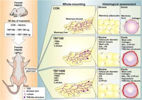
{"title":"Subacute tributyltin exposure alters the development and morphology of mammary glands in association with CYP19A1 expression in female rats","authors":"Natalia P. Silva , Charles S. da Costa , Kayke L. Barbosa , Cidália de F. Januario , Leticia N. Gama-de-Souza , Cinthia Breves , Rodrigo S. Fortunato , Leandro Miranda-Alves , Miriane de Oliveira , Celia R. Nogueira , Jones B. Graceli","doi":"10.1016/j.reprotox.2024.108635","DOIUrl":null,"url":null,"abstract":"<div><p>Tributyltin (TBT) is an endocrine-disrupting chemical (EDC) related to reproductive dysfunctions. However, few studies have investigated the effects of TBT exposure on mammary gland development. Thus, we assessed whether subacute TBT exposure causes irregularities in mammary gland development. We administered TBT (100 and 1,000 ng/kg/day for 30 days) to female rats from postnatal day (PND) 25 to PND 55, and mammary gland development, morphology, inflammation, collagen deposition, and protein expression were evaluated. Abnormal mammary gland development was observed in both TBT groups. Specifically, TBT exposure reduced the number of terminal end buds (TEBs), type 1 (AB1) alveolar buds, and type 2 (AB2) alveolar buds. An increase in the lobule and differentiation (DF) 2 score was found in the mammary glands of TBT rats. TBT exposure increased mammary gland blood vessels, mast cell numbers, and collagen deposition. Additionally, both TBT rats exhibited intraductal hyperplasia and TEB-like structures. An increase in estrogen receptor alpha (ERα), progesterone receptor (PR), and cytochrome P450 family 19 subfamily A member 1 (CYP19A1) - positive cells was observed in the mammary glands of TBT rats. A strong negative correlation was observed between CYP19A1- positive cells and TEB number. In addition, CYP19A1 - positive cells were positively correlated with mammary gland TEB-like structure, ductal hyperplasia, inflammation, and collagen deposition. Thus, these data suggest that TBT exposure impairs mammary gland development through the modulation of CYP19A1 signaling pathways in female rats.</p></div>","PeriodicalId":21137,"journal":{"name":"Reproductive toxicology","volume":null,"pages":null},"PeriodicalIF":3.3000,"publicationDate":"2024-06-26","publicationTypes":"Journal Article","fieldsOfStudy":null,"isOpenAccess":false,"openAccessPdf":"","citationCount":"0","resultStr":null,"platform":"Semanticscholar","paperid":null,"PeriodicalName":"Reproductive toxicology","FirstCategoryId":"3","ListUrlMain":"https://www.sciencedirect.com/science/article/pii/S0890623824001023","RegionNum":4,"RegionCategory":"医学","ArticlePicture":[],"TitleCN":null,"AbstractTextCN":null,"PMCID":null,"EPubDate":"","PubModel":"","JCR":"Q2","JCRName":"REPRODUCTIVE BIOLOGY","Score":null,"Total":0}
引用次数: 0
Abstract
Tributyltin (TBT) is an endocrine-disrupting chemical (EDC) related to reproductive dysfunctions. However, few studies have investigated the effects of TBT exposure on mammary gland development. Thus, we assessed whether subacute TBT exposure causes irregularities in mammary gland development. We administered TBT (100 and 1,000 ng/kg/day for 30 days) to female rats from postnatal day (PND) 25 to PND 55, and mammary gland development, morphology, inflammation, collagen deposition, and protein expression were evaluated. Abnormal mammary gland development was observed in both TBT groups. Specifically, TBT exposure reduced the number of terminal end buds (TEBs), type 1 (AB1) alveolar buds, and type 2 (AB2) alveolar buds. An increase in the lobule and differentiation (DF) 2 score was found in the mammary glands of TBT rats. TBT exposure increased mammary gland blood vessels, mast cell numbers, and collagen deposition. Additionally, both TBT rats exhibited intraductal hyperplasia and TEB-like structures. An increase in estrogen receptor alpha (ERα), progesterone receptor (PR), and cytochrome P450 family 19 subfamily A member 1 (CYP19A1) - positive cells was observed in the mammary glands of TBT rats. A strong negative correlation was observed between CYP19A1- positive cells and TEB number. In addition, CYP19A1 - positive cells were positively correlated with mammary gland TEB-like structure, ductal hyperplasia, inflammation, and collagen deposition. Thus, these data suggest that TBT exposure impairs mammary gland development through the modulation of CYP19A1 signaling pathways in female rats.
期刊介绍:
Drawing from a large number of disciplines, Reproductive Toxicology publishes timely, original research on the influence of chemical and physical agents on reproduction. Written by and for obstetricians, pediatricians, embryologists, teratologists, geneticists, toxicologists, andrologists, and others interested in detecting potential reproductive hazards, the journal is a forum for communication among researchers and practitioners. Articles focus on the application of in vitro, animal and clinical research to the practice of clinical medicine.
All aspects of reproduction are within the scope of Reproductive Toxicology, including the formation and maturation of male and female gametes, sexual function, the events surrounding the fusion of gametes and the development of the fertilized ovum, nourishment and transport of the conceptus within the genital tract, implantation, embryogenesis, intrauterine growth, placentation and placental function, parturition, lactation and neonatal survival. Adverse reproductive effects in males will be considered as significant as adverse effects occurring in females. To provide a balanced presentation of approaches, equal emphasis will be given to clinical and animal or in vitro work. Typical end points that will be studied by contributors include infertility, sexual dysfunction, spontaneous abortion, malformations, abnormal histogenesis, stillbirth, intrauterine growth retardation, prematurity, behavioral abnormalities, and perinatal mortality.
 求助内容:
求助内容: 应助结果提醒方式:


