{"title":"Extracellular vesicles in cattle infected with bovine leukaemia virus: isolation and molecular analysis.","authors":"Maria Szczotka, Magdalena Wasiak, Jacek Kuźmak","doi":"10.2478/jvetres-2024-0031","DOIUrl":null,"url":null,"abstract":"<p><strong>Introduction: </strong>Exosomes are nanosized lipid bilayer membranous microvesicles, extracellularly released from a variety of mammalian cells. They mediate intercellular signalling by transporting several types of RNA, lipids and proteins and participate in the intercellular exchange of DNA, RNA, micro RNA, proteins and other components. These microvesicles are present in all body fluids in physiological and pathological conditions and reflect the state of the host organism. The aim of the study was the isolation and molecular determination of exosomes in blood and supernatant fluids of bovine dendritic cell cultures infected with bovine leukaemia virus (BLV).</p><p><strong>Material and methods: </strong>Exosomes were isolated by ultracentrifugation from the blood sera, plasma and supernatant of bovine BLV-infected and uninfected control dendritic cell cultures and their presence was confirmed with scanning electron and transmission electron microscopy. Western blot analysis of the structural BLV glycoprotein 51 (Env) and protein 24 (Gag) and of the tetraspanin exosomal markers CD9, CD63 and flotillin-1 was undertaken in BLV+ and control BLV- cattle.</p><p><strong>Results: </strong>In exosomes of leukaemic cattle both BLV proteins and exosomal markers were detected. In healthy control animals only exosomal markers were determined.</p><p><strong>Conclusion: </strong>Proteins of BLV were released with exosomes and could be transferred into recipient cells as an alternative propagation route not requiring virus infection.</p>","PeriodicalId":17617,"journal":{"name":"Journal of Veterinary Research","volume":"68 2","pages":"189-198"},"PeriodicalIF":1.5000,"publicationDate":"2024-05-27","publicationTypes":"Journal Article","fieldsOfStudy":null,"isOpenAccess":false,"openAccessPdf":"https://www.ncbi.nlm.nih.gov/pmc/articles/PMC11210360/pdf/","citationCount":"0","resultStr":null,"platform":"Semanticscholar","paperid":null,"PeriodicalName":"Journal of Veterinary Research","FirstCategoryId":"97","ListUrlMain":"https://doi.org/10.2478/jvetres-2024-0031","RegionNum":3,"RegionCategory":"农林科学","ArticlePicture":[],"TitleCN":null,"AbstractTextCN":null,"PMCID":null,"EPubDate":"2024/6/1 0:00:00","PubModel":"eCollection","JCR":"Q2","JCRName":"VETERINARY SCIENCES","Score":null,"Total":0}
引用次数: 0
Abstract
Introduction: Exosomes are nanosized lipid bilayer membranous microvesicles, extracellularly released from a variety of mammalian cells. They mediate intercellular signalling by transporting several types of RNA, lipids and proteins and participate in the intercellular exchange of DNA, RNA, micro RNA, proteins and other components. These microvesicles are present in all body fluids in physiological and pathological conditions and reflect the state of the host organism. The aim of the study was the isolation and molecular determination of exosomes in blood and supernatant fluids of bovine dendritic cell cultures infected with bovine leukaemia virus (BLV).
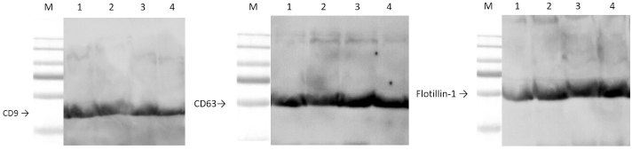
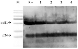
Material and methods: Exosomes were isolated by ultracentrifugation from the blood sera, plasma and supernatant of bovine BLV-infected and uninfected control dendritic cell cultures and their presence was confirmed with scanning electron and transmission electron microscopy. Western blot analysis of the structural BLV glycoprotein 51 (Env) and protein 24 (Gag) and of the tetraspanin exosomal markers CD9, CD63 and flotillin-1 was undertaken in BLV+ and control BLV- cattle.
Results: In exosomes of leukaemic cattle both BLV proteins and exosomal markers were detected. In healthy control animals only exosomal markers were determined.
Conclusion: Proteins of BLV were released with exosomes and could be transferred into recipient cells as an alternative propagation route not requiring virus infection.
期刊介绍:
Journal of Veterinary Research (formerly Bulletin of the Veterinary Institute in Pulawy) is a quarterly that publishes original papers, review articles and short communications on bacteriology, virology, parasitology, immunology, molecular biology, pathology, toxicology, pharmacology, and biochemistry. The main emphasis is, however, on infectious diseases of animals, food safety and public health, and clinical sciences.
 求助内容:
求助内容: 应助结果提醒方式:


