Zhou Liu, Peter Julius, Victor Mudenda, Guobin Kang, Luis Del Valle, John T West, Charles Wood
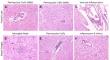
{"title":"Limited HIV-associated neuropathologies and lack of immune activation in sub-saharan African individuals with late-stage subtype C HIV-1 infection.","authors":"Zhou Liu, Peter Julius, Victor Mudenda, Guobin Kang, Luis Del Valle, John T West, Charles Wood","doi":"10.1007/s13365-024-01219-6","DOIUrl":null,"url":null,"abstract":"<p><p>Although previous studies have suggested that subtype B HIV-1 proviruses in the brain are associated with physiological changes and immune activation accompanied with microgliosis and astrogliosis, and indicated that both HIV-1 subtype variation and geographical location might influence the neuropathogenicity of HIV-1 in the brain. The natural course of neuropathogenesis of the most widespread subtype C HIV-1 has not been adequately investigated, especially for people living with HIV (PLWH) in sub-Saharan Africa. To characterize the natural neuropathology of subtype C HIV-1, postmortem frontal lobe and basal ganglia tissues were collected from nine ART-naïve individuals who died of late-stage AIDS with subtype C HIV-1 infection, and eight uninfected deceased individuals as controls. Histological staining was performed on all brain tissues to assess brain pathologies. Immunohistochemistry (IHC) against CD4, p24, Iba-1, GFAP, and CD8 in all brain tissues was conducted to evaluate potential viral production and immune activation. Histological results showed mild perivascular cuffs of lymphocytes only in a minority of the infected individuals. Viral capsid p24 protein was only detected in circulating immune cells of one infected individual, suggesting a lack of productive HIV-1 infection of the brain even at the late-stage of AIDS. Notably, similar levels of Iba-1 or GFAP between HIV + and HIV- brain tissues indicated a lack of microgliosis and astrogliosis, respectively. Similar levels of CD8 + cytotoxic T lymphocyte (CTL) infiltration between HIV + and HIV- brain tissues indicated CTL were not likely to be involved within subtype C HIV-1 infected participants of this cohort. Results from this subtype C HIV-1 study suggest that there is a lack of productive infection and limited neuropathogenesis by subtype C HIV-1 even at late-stage disease, which is in contrast to what was reported for subtype B HIV-1 by other investigators.</p>","PeriodicalId":16665,"journal":{"name":"Journal of NeuroVirology","volume":" ","pages":"303-315"},"PeriodicalIF":1.9000,"publicationDate":"2024-06-01","publicationTypes":"Journal Article","fieldsOfStudy":null,"isOpenAccess":false,"openAccessPdf":"","citationCount":"0","resultStr":null,"platform":"Semanticscholar","paperid":null,"PeriodicalName":"Journal of NeuroVirology","FirstCategoryId":"3","ListUrlMain":"https://doi.org/10.1007/s13365-024-01219-6","RegionNum":4,"RegionCategory":"医学","ArticlePicture":[],"TitleCN":null,"AbstractTextCN":null,"PMCID":null,"EPubDate":"2024/6/28 0:00:00","PubModel":"Epub","JCR":"Q3","JCRName":"NEUROSCIENCES","Score":null,"Total":0}
引用次数: 0
Abstract
Although previous studies have suggested that subtype B HIV-1 proviruses in the brain are associated with physiological changes and immune activation accompanied with microgliosis and astrogliosis, and indicated that both HIV-1 subtype variation and geographical location might influence the neuropathogenicity of HIV-1 in the brain. The natural course of neuropathogenesis of the most widespread subtype C HIV-1 has not been adequately investigated, especially for people living with HIV (PLWH) in sub-Saharan Africa. To characterize the natural neuropathology of subtype C HIV-1, postmortem frontal lobe and basal ganglia tissues were collected from nine ART-naïve individuals who died of late-stage AIDS with subtype C HIV-1 infection, and eight uninfected deceased individuals as controls. Histological staining was performed on all brain tissues to assess brain pathologies. Immunohistochemistry (IHC) against CD4, p24, Iba-1, GFAP, and CD8 in all brain tissues was conducted to evaluate potential viral production and immune activation. Histological results showed mild perivascular cuffs of lymphocytes only in a minority of the infected individuals. Viral capsid p24 protein was only detected in circulating immune cells of one infected individual, suggesting a lack of productive HIV-1 infection of the brain even at the late-stage of AIDS. Notably, similar levels of Iba-1 or GFAP between HIV + and HIV- brain tissues indicated a lack of microgliosis and astrogliosis, respectively. Similar levels of CD8 + cytotoxic T lymphocyte (CTL) infiltration between HIV + and HIV- brain tissues indicated CTL were not likely to be involved within subtype C HIV-1 infected participants of this cohort. Results from this subtype C HIV-1 study suggest that there is a lack of productive infection and limited neuropathogenesis by subtype C HIV-1 even at late-stage disease, which is in contrast to what was reported for subtype B HIV-1 by other investigators.
期刊介绍:
The Journal of NeuroVirology (JNV) provides a unique platform for the publication of high-quality basic science and clinical studies on the molecular biology and pathogenesis of viral infections of the nervous system, and for reporting on the development of novel therapeutic strategies using neurotropic viral vectors. The Journal also emphasizes publication of non-viral infections that affect the central nervous system. The Journal publishes original research articles, reviews, case reports, coverage of various scientific meetings, along with supplements and special issues on selected subjects.
The Journal is currently accepting submissions of original work from the following basic and clinical research areas: Aging & Neurodegeneration, Apoptosis, CNS Signal Transduction, Emerging CNS Infections, Molecular Virology, Neural-Immune Interaction, Novel Diagnostics, Novel Therapeutics, Stem Cell Biology, Transmissable Encephalopathies/Prion, Vaccine Development, Viral Genomics, Viral Neurooncology, Viral Neurochemistry, Viral Neuroimmunology, Viral Neuropharmacology.
 求助内容:
求助内容: 应助结果提醒方式:


