Guillaume Cogan MD, Kensuke Daida MD, PhD, Kimberley J. Billingsley PhD, Christelle Tesson PhD, Sylvie Forlani PhD, Ludmila Jornea MSc, Lionel Arnaud PhD, Laurène Tissier MLT, Eric LeGuern MD, PhD, Andrew B. Singleton PhD, Mélanie Ferrien MSc, Hélène Gervais Bernard MD, Suzanne Lesage PhD, Cornelis Blauwendraat PhD, Alexis Brice MD
{"title":"Long-Read Sequencing Unravels the Complexity of Structural Variants in PRKN in Two Individuals with Early-Onset Parkinson's Disease","authors":"Guillaume Cogan MD, Kensuke Daida MD, PhD, Kimberley J. Billingsley PhD, Christelle Tesson PhD, Sylvie Forlani PhD, Ludmila Jornea MSc, Lionel Arnaud PhD, Laurène Tissier MLT, Eric LeGuern MD, PhD, Andrew B. Singleton PhD, Mélanie Ferrien MSc, Hélène Gervais Bernard MD, Suzanne Lesage PhD, Cornelis Blauwendraat PhD, Alexis Brice MD","doi":"10.1002/mds.29914","DOIUrl":null,"url":null,"abstract":"<p>About 5% to 10% of Parkinson's disease (PD) cases are monogenic; otherwise PD is generally known to be idiopathic. Although more than a dozen genes that contain disease-causing mutations have been identified to date, <i>PRKN</i> is the most frequently mutated gene in autosomal recessive early-onset PD (EOPD).<span><sup>1</sup></span> However, the genetic cause of patients with a typical <i>PRKN</i> phenotype is sometimes elusive because of the limitations of traditional genetic methods to detect complex structural mutations that are frequent in <i>PRKN</i>.<span><sup>2</sup></span></p><p>The phenotype is usually specific, consisting of a slowly progressive EOPD with a good and long-standing response to levodopa. Dystonia, dyskinesia, and motor fluctuations are typical, whereas autonomic dysfunction, psychotic symptoms, and cognitive decline are usually absent.<span><sup>3</sup></span> We report 2 siblings of European ancestries exhibiting <i>PRKN</i> phenotype left undiagnosed for years after multiple genetic investigations (Fig. 1).</p><p>Siblings II-2 and II-4 presented at age 31 and 33 years, respectively, with asymmetrical limb akinesia associated with resting tremor with no medical history and no parental consanguinity. Cerebral magnetic resonance imaging was normal, and Wilson's disease biomarkers were negative. Focal and paroxysmal dystonia was present in II-4. The disease slowly evolved with a low <i>off</i>-medication state UPDRS (Unified Parkinson's Disease Rating Scale) 13 and 16 years after disease onset (scores of 33 and 35 for II-2 and II-4, respectively). Initial response to levodopa was remarkable for both (90% and 80%). At last examination, II-4 had dyskinesia and motor fluctuations. Of note, at the most recent examination (age 45 and 47 years), cognitive impairment, postural instability, neurogenic bladder, and bowel dysfunction were absent.</p><p>Because this presentation was consistent with <i>PRKN</i>-PD, we first performed <i>PRKN</i> multiple ligation probe amplification (MLPA) and Sanger sequencing, which revealed one copy of exon 4 for both individuals and the absence of pathogenic single-nucleotide variant, interpreted as a heterozygous exon 4 deletion (Fig. S1). Multiple genetic investigations, including another MLPA, digital droplet polymerase chain reaction, and targeted and exome sequencing, confirmed the presence of one copy of exon 4, without any additional pathogenic variant. Thus, this result was not sufficient to explain the phenotype.</p><p>Next we performed Oxford Nanopore long-read sequencing (LRS) for one individual using a protocol reported previously (https://www.protocols.io/view/processing-frozen-cells-for-population-scale-sqk-l-6qpvr347bvmk/v1). LRS detected a large compound heterozygous 178-kb deletion and 106-kb duplication, encompassing exons 3 and 4 and exon 3, respectively (Fig. 1). Both DNA loss and gain of the same exons 3 and 4 are described in typical <i>PRKN</i>-PD individuals, as reported in the movement disorders society gene database (https://www.mdsgene.org/d/1/g/4). Breakpoint junction PCR confirmed the presence of the two structural variants and revealed both variants in the second individual (Fig. S2). LRS did not identify any additional variants in PD known genes. Because both deletion and duplication breakpoints were located in deep intronic regions and genetic dosage of exon 3 was normal, short-read sequencing and other methods could not detect the complex and balanced rearrangement. Overall, these results demonstrated that biallelic <i>PRKN</i> variants were the cause of PD in this family.</p><p>As shown by a previous study, we here confirm the potential of LRS to determine complex <i>PRKN</i> structural variants in unsolved <i>PRKN-</i>PD cases.<span><sup>4</sup></span></p><p>G.C. is supported by the Global Parkinson's Genetics Program (GP2). GP2 is funded by the Aligning Science Across Parkinson's (ASAP) initiative and implemented by The Michael J. Fox Foundation for Parkinson's Research (https://gp2.org). For a complete list of GP2 members see https://gp2.org. K.D. reports receiving grants from the JSPS Research Fellowship for Japanese Biomedical and Behavioral Researchers at NIH. S.L. has received grants from <i>Fondation de la Recherche Médicale</i> (FRM, MND202004011718).</p><p>G.C: Design, execution, analysis, writing and editing of final version.</p><p>K.D: Analysis and editing of final version.</p><p>K.J.B: Execution.</p><p>C.T: Execution, analysis.</p><p>S.F: Execution.</p><p>L.J: Execution.</p><p>L.A: Execution, analysis and editing of final version.</p><p>L.T: Execution.</p><p>EL: Execution.</p><p>A.B.S: Design and editing of final version.</p><p>M.F: Execution.</p><p>H.G.B: Execution.</p><p>S.L: Execution.</p><p>C.B: Design and editing of final version.</p><p>A.B: Design, writing and editing of final version.</p>","PeriodicalId":213,"journal":{"name":"Movement Disorders","volume":null,"pages":null},"PeriodicalIF":7.4000,"publicationDate":"2024-06-28","publicationTypes":"Journal Article","fieldsOfStudy":null,"isOpenAccess":false,"openAccessPdf":"https://onlinelibrary.wiley.com/doi/epdf/10.1002/mds.29914","citationCount":"0","resultStr":null,"platform":"Semanticscholar","paperid":null,"PeriodicalName":"Movement Disorders","FirstCategoryId":"3","ListUrlMain":"https://onlinelibrary.wiley.com/doi/10.1002/mds.29914","RegionNum":1,"RegionCategory":"医学","ArticlePicture":[],"TitleCN":null,"AbstractTextCN":null,"PMCID":null,"EPubDate":"","PubModel":"","JCR":"Q1","JCRName":"CLINICAL NEUROLOGY","Score":null,"Total":0}
引用次数: 0
Abstract
About 5% to 10% of Parkinson's disease (PD) cases are monogenic; otherwise PD is generally known to be idiopathic. Although more than a dozen genes that contain disease-causing mutations have been identified to date, PRKN is the most frequently mutated gene in autosomal recessive early-onset PD (EOPD).1 However, the genetic cause of patients with a typical PRKN phenotype is sometimes elusive because of the limitations of traditional genetic methods to detect complex structural mutations that are frequent in PRKN.2
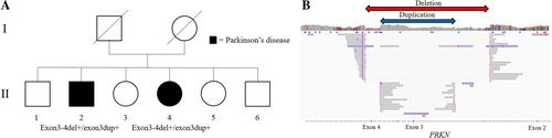
The phenotype is usually specific, consisting of a slowly progressive EOPD with a good and long-standing response to levodopa. Dystonia, dyskinesia, and motor fluctuations are typical, whereas autonomic dysfunction, psychotic symptoms, and cognitive decline are usually absent.3 We report 2 siblings of European ancestries exhibiting PRKN phenotype left undiagnosed for years after multiple genetic investigations (Fig. 1).
Siblings II-2 and II-4 presented at age 31 and 33 years, respectively, with asymmetrical limb akinesia associated with resting tremor with no medical history and no parental consanguinity. Cerebral magnetic resonance imaging was normal, and Wilson's disease biomarkers were negative. Focal and paroxysmal dystonia was present in II-4. The disease slowly evolved with a low off-medication state UPDRS (Unified Parkinson's Disease Rating Scale) 13 and 16 years after disease onset (scores of 33 and 35 for II-2 and II-4, respectively). Initial response to levodopa was remarkable for both (90% and 80%). At last examination, II-4 had dyskinesia and motor fluctuations. Of note, at the most recent examination (age 45 and 47 years), cognitive impairment, postural instability, neurogenic bladder, and bowel dysfunction were absent.
Because this presentation was consistent with PRKN-PD, we first performed PRKN multiple ligation probe amplification (MLPA) and Sanger sequencing, which revealed one copy of exon 4 for both individuals and the absence of pathogenic single-nucleotide variant, interpreted as a heterozygous exon 4 deletion (Fig. S1). Multiple genetic investigations, including another MLPA, digital droplet polymerase chain reaction, and targeted and exome sequencing, confirmed the presence of one copy of exon 4, without any additional pathogenic variant. Thus, this result was not sufficient to explain the phenotype.
Next we performed Oxford Nanopore long-read sequencing (LRS) for one individual using a protocol reported previously (https://www.protocols.io/view/processing-frozen-cells-for-population-scale-sqk-l-6qpvr347bvmk/v1). LRS detected a large compound heterozygous 178-kb deletion and 106-kb duplication, encompassing exons 3 and 4 and exon 3, respectively (Fig. 1). Both DNA loss and gain of the same exons 3 and 4 are described in typical PRKN-PD individuals, as reported in the movement disorders society gene database (https://www.mdsgene.org/d/1/g/4). Breakpoint junction PCR confirmed the presence of the two structural variants and revealed both variants in the second individual (Fig. S2). LRS did not identify any additional variants in PD known genes. Because both deletion and duplication breakpoints were located in deep intronic regions and genetic dosage of exon 3 was normal, short-read sequencing and other methods could not detect the complex and balanced rearrangement. Overall, these results demonstrated that biallelic PRKN variants were the cause of PD in this family.
As shown by a previous study, we here confirm the potential of LRS to determine complex PRKN structural variants in unsolved PRKN-PD cases.4
G.C. is supported by the Global Parkinson's Genetics Program (GP2). GP2 is funded by the Aligning Science Across Parkinson's (ASAP) initiative and implemented by The Michael J. Fox Foundation for Parkinson's Research (https://gp2.org). For a complete list of GP2 members see https://gp2.org. K.D. reports receiving grants from the JSPS Research Fellowship for Japanese Biomedical and Behavioral Researchers at NIH. S.L. has received grants from Fondation de la Recherche Médicale (FRM, MND202004011718).
G.C: Design, execution, analysis, writing and editing of final version.
K.D: Analysis and editing of final version.
K.J.B: Execution.
C.T: Execution, analysis.
S.F: Execution.
L.J: Execution.
L.A: Execution, analysis and editing of final version.
L.T: Execution.
EL: Execution.
A.B.S: Design and editing of final version.
M.F: Execution.
H.G.B: Execution.
S.L: Execution.
C.B: Design and editing of final version.
A.B: Design, writing and editing of final version.
期刊介绍:
Movement Disorders publishes a variety of content types including Reviews, Viewpoints, Full Length Articles, Historical Reports, Brief Reports, and Letters. The journal considers original manuscripts on topics related to the diagnosis, therapeutics, pharmacology, biochemistry, physiology, etiology, genetics, and epidemiology of movement disorders. Appropriate topics include Parkinsonism, Chorea, Tremors, Dystonia, Myoclonus, Tics, Tardive Dyskinesia, Spasticity, and Ataxia.

 求助内容:
求助内容: 应助结果提醒方式:


