Elena B. Kiseleva, Alexander A. Sovetsky, Maksim G. Ryabkov, Ekaterina V. Gubarkova, Anton A. Plekhanov, Evgeniya L. Bederina, Arseniy L. Potapov, Alexandra Y. Bogomolova, Vladimir Y. Zaitsev, Natalia D. Gladkova
{"title":"Detecting emergence of ruptures in individual layers of the stretched intestinal wall using optical coherence elastography: A pilot study","authors":"Elena B. Kiseleva, Alexander A. Sovetsky, Maksim G. Ryabkov, Ekaterina V. Gubarkova, Anton A. Plekhanov, Evgeniya L. Bederina, Arseniy L. Potapov, Alexandra Y. Bogomolova, Vladimir Y. Zaitsev, Natalia D. Gladkova","doi":"10.1002/jbio.202400086","DOIUrl":null,"url":null,"abstract":"<p>We report a new application of compression optical coherence elastography (C-OCE) to monitor the emergence of ruptures in individual layers of longitudinally stretched small-intestine walls using tissue samples (<i>n</i> = 36) from nine minipigs. Before stretching, C-OCE successfully estimated stiffness for each intestine-wall layer: longitudinal muscular layer with serosa, circumferential muscular layer, submucosa and mucosa. In stretched samples, C-OCE clearly visualized initial stiffening in both muscular layers. By 25% elongation, a sharp stiffness decrease for the longitudinal muscular layer, indicated emergence of tears in all samples. With further stretching, for most samples, ruptures emerged in the circumferential muscular layer and submucosa, while mucosa remained undamaged. Histology confirmed the OCE-revealed damaging and absence of tissue damage for ~15% elongation. Thus, C-OCE has demonstrated a high potential for determining the safety tissue-stretching threshold which afterward may be used intraoperatively to prevent rupture risk in intestinal tissues stretched during various diagnostic/therapeutic procedures.</p>","PeriodicalId":184,"journal":{"name":"Journal of Biophotonics","volume":"17 8","pages":""},"PeriodicalIF":2.0000,"publicationDate":"2024-06-25","publicationTypes":"Journal Article","fieldsOfStudy":null,"isOpenAccess":false,"openAccessPdf":"","citationCount":"0","resultStr":null,"platform":"Semanticscholar","paperid":null,"PeriodicalName":"Journal of Biophotonics","FirstCategoryId":"101","ListUrlMain":"https://onlinelibrary.wiley.com/doi/10.1002/jbio.202400086","RegionNum":3,"RegionCategory":"物理与天体物理","ArticlePicture":[],"TitleCN":null,"AbstractTextCN":null,"PMCID":null,"EPubDate":"","PubModel":"","JCR":"Q3","JCRName":"BIOCHEMICAL RESEARCH METHODS","Score":null,"Total":0}
引用次数: 0
Abstract
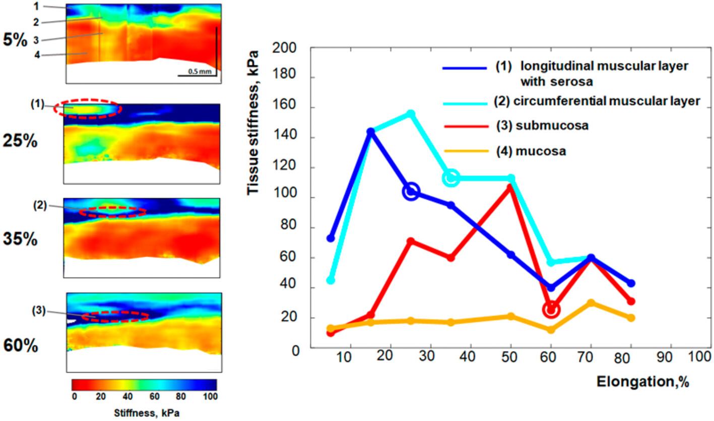
We report a new application of compression optical coherence elastography (C-OCE) to monitor the emergence of ruptures in individual layers of longitudinally stretched small-intestine walls using tissue samples (n = 36) from nine minipigs. Before stretching, C-OCE successfully estimated stiffness for each intestine-wall layer: longitudinal muscular layer with serosa, circumferential muscular layer, submucosa and mucosa. In stretched samples, C-OCE clearly visualized initial stiffening in both muscular layers. By 25% elongation, a sharp stiffness decrease for the longitudinal muscular layer, indicated emergence of tears in all samples. With further stretching, for most samples, ruptures emerged in the circumferential muscular layer and submucosa, while mucosa remained undamaged. Histology confirmed the OCE-revealed damaging and absence of tissue damage for ~15% elongation. Thus, C-OCE has demonstrated a high potential for determining the safety tissue-stretching threshold which afterward may be used intraoperatively to prevent rupture risk in intestinal tissues stretched during various diagnostic/therapeutic procedures.
期刊介绍:
The first international journal dedicated to publishing reviews and original articles from this exciting field, the Journal of Biophotonics covers the broad range of research on interactions between light and biological material. The journal offers a platform where the physicist communicates with the biologist and where the clinical practitioner learns about the latest tools for the diagnosis of diseases. As such, the journal is highly interdisciplinary, publishing cutting edge research in the fields of life sciences, medicine, physics, chemistry, and engineering. The coverage extends from fundamental research to specific developments, while also including the latest applications.
 求助内容:
求助内容: 应助结果提醒方式:


