Pial collaterals limit stroke progression and metabolic stress in hypoperfused tissue: An MRI perfusion and mq-BOLD study

IF 2.3 4区 医学 Q3 CLINICAL NEUROLOGY
Franziska Frank, Stephan Kaczmarz, Christine Preibisch, Ralf Deichmann, Ulrike Nöth, Marlies Wagner, Alexander Seiler
{"title":"Pial collaterals limit stroke progression and metabolic stress in hypoperfused tissue: An MRI perfusion and mq-BOLD study","authors":"Franziska Frank,&nbsp;Stephan Kaczmarz,&nbsp;Christine Preibisch,&nbsp;Ralf Deichmann,&nbsp;Ulrike Nöth,&nbsp;Marlies Wagner,&nbsp;Alexander Seiler","doi":"10.1111/jon.13220","DOIUrl":null,"url":null,"abstract":"<div>\n \n \n <section>\n \n <h3> Background and Purpose</h3>\n \n <p>In acute ischemic stroke (AIS) due to large-vessel occlusion (LVO), the relationship between cerebral oxygen extraction fraction (OEF) as the hallmark of the ischemic penumbra and leptomeningeal collateral supply is not well established. We aimed to investigate the relationship between pial collateralization and tissue oxygen extraction in patients with LVO using magnetic resonance imaging (MRI).</p>\n </section>\n \n <section>\n \n <h3> Methods</h3>\n \n <p>Data from 14 patients with anterior circulation LVO who underwent MRI before acute stroke treatment were analyzed. In addition to diffusion-weighted imaging and perfusion-weighted imaging (PWI), the protocol comprised sequences for multiparametric quantitative blood-oxygen-level-dependent imaging for the calculation of relative OEF (rOEF). Pial collateral supply was quantitatively assessed by analyzing the signal variance in T2*-weighted PWI time series. Relationships between collateral supply, infarct volume, rOEF in peri-infarct hypoperfused tissue, and clinical stroke severity were assessed.</p>\n </section>\n \n <section>\n \n <h3> Results</h3>\n \n <p>The PWI-based parameter quantifying collateral supply was negatively correlated with baseline ischemic core volume and rOEF in the hypoperfused peri-infarct area (<i>p</i> &lt; .01). Both reduced collateral supply and increased rOEF correlated significantly with higher scores on the National Institutes of Health Stroke Scale (<i>p</i> &lt; .05). Increased rOEF within hypoperfused tissue was associated with higher baseline (<i>p</i> = .043) and follow-up infarct volume (<i>p</i> = .009).</p>\n </section>\n \n <section>\n \n <h3> Conclusions</h3>\n \n <p>Signal variance-based mapping of collaterals with PWI depicts pial collateral supply, which is closely tied to tissue pathophysiology and clinical and imaging outcomes. Magnetic-resonance-derived mapping of cerebral rOEF reveals penumbral characteristics of hypoperfused tissue and might provide a promising imaging biomarker in AIS.</p>\n </section>\n </div>","PeriodicalId":16399,"journal":{"name":"Journal of Neuroimaging","volume":"34 5","pages":"562-566"},"PeriodicalIF":2.3000,"publicationDate":"2024-06-26","publicationTypes":"Journal Article","fieldsOfStudy":null,"isOpenAccess":false,"openAccessPdf":"https://onlinelibrary.wiley.com/doi/epdf/10.1111/jon.13220","citationCount":"0","resultStr":null,"platform":"Semanticscholar","paperid":null,"PeriodicalName":"Journal of Neuroimaging","FirstCategoryId":"3","ListUrlMain":"https://onlinelibrary.wiley.com/doi/10.1111/jon.13220","RegionNum":4,"RegionCategory":"医学","ArticlePicture":[],"TitleCN":null,"AbstractTextCN":null,"PMCID":null,"EPubDate":"","PubModel":"","JCR":"Q3","JCRName":"CLINICAL NEUROLOGY","Score":null,"Total":0}
引用次数: 0

Abstract

Background and Purpose

In acute ischemic stroke (AIS) due to large-vessel occlusion (LVO), the relationship between cerebral oxygen extraction fraction (OEF) as the hallmark of the ischemic penumbra and leptomeningeal collateral supply is not well established. We aimed to investigate the relationship between pial collateralization and tissue oxygen extraction in patients with LVO using magnetic resonance imaging (MRI).

Methods

Data from 14 patients with anterior circulation LVO who underwent MRI before acute stroke treatment were analyzed. In addition to diffusion-weighted imaging and perfusion-weighted imaging (PWI), the protocol comprised sequences for multiparametric quantitative blood-oxygen-level-dependent imaging for the calculation of relative OEF (rOEF). Pial collateral supply was quantitatively assessed by analyzing the signal variance in T2*-weighted PWI time series. Relationships between collateral supply, infarct volume, rOEF in peri-infarct hypoperfused tissue, and clinical stroke severity were assessed.

Results

The PWI-based parameter quantifying collateral supply was negatively correlated with baseline ischemic core volume and rOEF in the hypoperfused peri-infarct area (p < .01). Both reduced collateral supply and increased rOEF correlated significantly with higher scores on the National Institutes of Health Stroke Scale (p < .05). Increased rOEF within hypoperfused tissue was associated with higher baseline (p = .043) and follow-up infarct volume (p = .009).

Conclusions

Signal variance-based mapping of collaterals with PWI depicts pial collateral supply, which is closely tied to tissue pathophysiology and clinical and imaging outcomes. Magnetic-resonance-derived mapping of cerebral rOEF reveals penumbral characteristics of hypoperfused tissue and might provide a promising imaging biomarker in AIS.
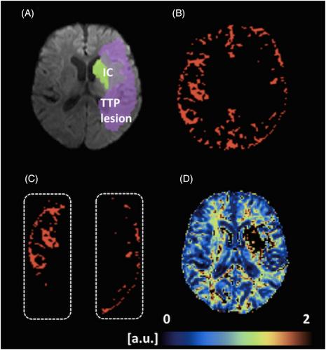
Abstract Image

在低灌注组织中,静脉瓣限制了中风的进展和代谢压力:磁共振成像灌注和 mq-BOLD 研究。
背景和目的:在大血管闭塞(LVO)导致的急性缺血性卒中(AIS)中,作为缺血半影标志的脑氧萃取率(OEF)与脑侧膜侧供之间的关系尚未明确。我们的目的是利用磁共振成像(MRI)研究 LVO 患者的静脉侧支与组织氧萃取之间的关系:方法:分析了 14 名前循环 LVO 患者的数据,这些患者在急性卒中治疗前接受了 MRI 检查。除了弥散加权成像和灌注加权成像(PWI)外,方案还包括用于计算相对 OEF(rOEF)的多参数定量血氧水平依赖性成像序列。通过分析 T2* 加权 PWI 时间序列中的信号方差,对皮质侧支供应进行定量评估。评估了侧支供应、梗死容积、梗死周围低灌注组织的 rOEF 和临床卒中严重程度之间的关系:结果:基于脉搏波成像的侧支供应量化参数与基线缺血核心容积和梗死周围低灌注区的 rOEF 呈负相关(p 结论:基于脉搏波成像的侧支供应量化参数与基线缺血核心容积和梗死周围低灌注区的 rOEF 呈负相关(p 结论):基于信号方差的脉搏波成像侧支映射描述了与组织病理生理学、临床和成像结果密切相关的侧支供应。磁共振衍生的大脑 rOEF 映射揭示了低灌注组织的半影特征,可能为 AIS 提供一种有前景的成像生物标志物。
本文章由计算机程序翻译,如有差异,请以英文原文为准。
求助全文
约1分钟内获得全文 求助全文
来源期刊
Journal of Neuroimaging
Journal of Neuroimaging 医学-核医学
CiteScore
4.70
自引率
0.00%
发文量
117
审稿时长
6-12 weeks
期刊介绍: Start reading the Journal of Neuroimaging to learn the latest neurological imaging techniques. The peer-reviewed research is written in a practical clinical context, giving you the information you need on: MRI CT Carotid Ultrasound and TCD SPECT PET Endovascular Surgical Neuroradiology Functional MRI Xenon CT and other new and upcoming neuroscientific modalities.The Journal of Neuroimaging addresses the full spectrum of human nervous system disease, including stroke, neoplasia, degenerating and demyelinating disease, epilepsy, tumors, lesions, infectious disease, cerebral vascular arterial diseases, toxic-metabolic disease, psychoses, dementias, heredo-familial disease, and trauma.Offering original research, review articles, case reports, neuroimaging CPCs, and evaluations of instruments and technology relevant to the nervous system, the Journal of Neuroimaging focuses on useful clinical developments and applications, tested techniques and interpretations, patient care, diagnostics, and therapeutics. Start reading today!
×
引用
GB/T 7714-2015
复制
MLA
复制
APA
复制
导出至
BibTeX EndNote RefMan NoteFirst NoteExpress
×
提示
您的信息不完整,为了账户安全,请先补充。
现在去补充
×
提示
您因"违规操作"
具体请查看互助需知
我知道了
×
提示
确定
请完成安全验证×
copy
已复制链接
快去分享给好友吧!
我知道了
右上角分享
点击右上角分享
0
联系我们:info@booksci.cn Book学术提供免费学术资源搜索服务,方便国内外学者检索中英文文献。致力于提供最便捷和优质的服务体验。 Copyright © 2023 布克学术 All rights reserved.
京ICP备2023020795号-1
ghs 京公网安备 11010802042870号
Book学术文献互助
Book学术文献互助群
群 号:481959085
Book学术官方微信