{"title":"Compressive confocal microscopy imaging at the single-photon level with ultra-low sampling ratios","authors":"Shuai Liu, Bin Chen, Wenzhen Zou, Hao Sha, Xiaochen Feng, Sanyang Han, Xiu Li, Xuri Yao, Jian Zhang, Yongbing Zhang","doi":"10.1038/s44172-024-00236-x","DOIUrl":null,"url":null,"abstract":"Laser-scanning confocal microscopy serves as a critical instrument for microscopic research in biology. However, it suffers from low imaging speed and high phototoxicity. Here we build a novel deep compressive confocal microscope, which employs a digital micromirror device as a coding mask for single-pixel imaging and a pinhole for confocal microscopic imaging respectively. Combined with a deep learning reconstruction algorithm, our system is able to achieve high-quality confocal microscopic imaging with low phototoxicity. Our imaging experiments with fluorescent microspheres demonstrate its capability of achieving single-pixel confocal imaging with a sampling ratio of only approximately 0.03% in specific sparse scenarios. Moreover, the deep compressive confocal microscope allows single-pixel imaging at the single-photon level, thus reducing the excitation light power requirement for confocal imaging and suppressing the phototoxicity. We believe that our system has great potential for long-duration and high-speed microscopic imaging of living cells. Shuai Liu, Bin Chen and colleagues improve imaging speed and reduced phototoxicity in confocal microimaging by building a deep compressive confocal microscope. Digital micromirror acts as a coding mask for deep learning-based reconstruction algorithms.","PeriodicalId":72644,"journal":{"name":"Communications engineering","volume":" ","pages":"1-9"},"PeriodicalIF":0.0000,"publicationDate":"2024-06-25","publicationTypes":"Journal Article","fieldsOfStudy":null,"isOpenAccess":false,"openAccessPdf":"https://www.nature.com/articles/s44172-024-00236-x.pdf","citationCount":"0","resultStr":null,"platform":"Semanticscholar","paperid":null,"PeriodicalName":"Communications engineering","FirstCategoryId":"1085","ListUrlMain":"https://www.nature.com/articles/s44172-024-00236-x","RegionNum":0,"RegionCategory":null,"ArticlePicture":[],"TitleCN":null,"AbstractTextCN":null,"PMCID":null,"EPubDate":"","PubModel":"","JCR":"","JCRName":"","Score":null,"Total":0}
引用次数: 0
Abstract
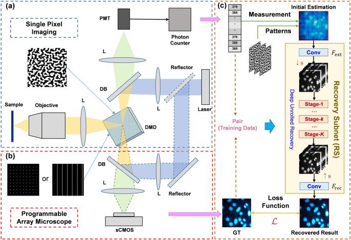
Laser-scanning confocal microscopy serves as a critical instrument for microscopic research in biology. However, it suffers from low imaging speed and high phototoxicity. Here we build a novel deep compressive confocal microscope, which employs a digital micromirror device as a coding mask for single-pixel imaging and a pinhole for confocal microscopic imaging respectively. Combined with a deep learning reconstruction algorithm, our system is able to achieve high-quality confocal microscopic imaging with low phototoxicity. Our imaging experiments with fluorescent microspheres demonstrate its capability of achieving single-pixel confocal imaging with a sampling ratio of only approximately 0.03% in specific sparse scenarios. Moreover, the deep compressive confocal microscope allows single-pixel imaging at the single-photon level, thus reducing the excitation light power requirement for confocal imaging and suppressing the phototoxicity. We believe that our system has great potential for long-duration and high-speed microscopic imaging of living cells. Shuai Liu, Bin Chen and colleagues improve imaging speed and reduced phototoxicity in confocal microimaging by building a deep compressive confocal microscope. Digital micromirror acts as a coding mask for deep learning-based reconstruction algorithms.

 求助内容:
求助内容: 应助结果提醒方式:


