{"title":"Loss of insulin-expressing extra-islet cells in type 1 diabetes is accompanied with increased number of glucagon-expressing extra-islet cells.","authors":"Louise Granlund, Marcus Lundberg","doi":"10.1007/s00428-024-03842-4","DOIUrl":null,"url":null,"abstract":"<p><p>The presence of remaining insulin-positive cells in type 1 diabetes (T1D) is well-known. These cells are part of islets or appear as extra-islet insulin-positive cells scattered in the exocrine parenchyma. The latter are poorly described, and the presence of scattered endocrine cells expressing other islet hormones than insulin has not been explored. This study aimed to compare the extra-islet insulin- or glucagon-positive cells concerning their frequency, transcription-factor expression, and mitotic activity in subjects with and without T1D. Multispectral imaging was used to examine extra-islet cells by staining for insulin, glucagon, ARX, PDX1, and Ki67. This was done in well-preserved pancreatic tissue obtained from heart-beating organ donors with or without T1D. In three T1D donors, lobes with insulin-containing islets (ICI) were found. Within these, a higher frequency of extra-islet insulin-positive cells was observed compared to lobes with insulin-deficient islets (IDI). Increased frequency of glucagon-positive extra-islet cells was observed in donors with T1D (median 53 cells/mm<sup>2</sup>) when compared with non-diabetic donors (11 cells/mm<sup>2</sup>, p = 0.004). Proliferating endocrine cells were present in donors with, and without T1D, as demonstrated by Ki67-positive staining (0-3% of the cells expressing insulin or glucagon). The reduced frequency of extra-islet insulin-positive cells in lobes with IDI in donors with T1D suggests that the pathological mechanism causing beta cell demise in T1D affects entire lobes. The presence of an increased frequency of glucagon-positive extra-islet cells supports the notion of a preserved capacity to regenerate the endocrine pancreas in donors with T1D.</p>","PeriodicalId":23514,"journal":{"name":"Virchows Archiv","volume":" ","pages":"687-695"},"PeriodicalIF":3.4000,"publicationDate":"2025-04-01","publicationTypes":"Journal Article","fieldsOfStudy":null,"isOpenAccess":false,"openAccessPdf":"https://www.ncbi.nlm.nih.gov/pmc/articles/PMC12018523/pdf/","citationCount":"0","resultStr":null,"platform":"Semanticscholar","paperid":null,"PeriodicalName":"Virchows Archiv","FirstCategoryId":"3","ListUrlMain":"https://doi.org/10.1007/s00428-024-03842-4","RegionNum":3,"RegionCategory":"医学","ArticlePicture":[],"TitleCN":null,"AbstractTextCN":null,"PMCID":null,"EPubDate":"2024/6/26 0:00:00","PubModel":"Epub","JCR":"Q1","JCRName":"PATHOLOGY","Score":null,"Total":0}
引用次数: 0
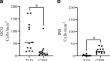
Abstract
The presence of remaining insulin-positive cells in type 1 diabetes (T1D) is well-known. These cells are part of islets or appear as extra-islet insulin-positive cells scattered in the exocrine parenchyma. The latter are poorly described, and the presence of scattered endocrine cells expressing other islet hormones than insulin has not been explored. This study aimed to compare the extra-islet insulin- or glucagon-positive cells concerning their frequency, transcription-factor expression, and mitotic activity in subjects with and without T1D. Multispectral imaging was used to examine extra-islet cells by staining for insulin, glucagon, ARX, PDX1, and Ki67. This was done in well-preserved pancreatic tissue obtained from heart-beating organ donors with or without T1D. In three T1D donors, lobes with insulin-containing islets (ICI) were found. Within these, a higher frequency of extra-islet insulin-positive cells was observed compared to lobes with insulin-deficient islets (IDI). Increased frequency of glucagon-positive extra-islet cells was observed in donors with T1D (median 53 cells/mm2) when compared with non-diabetic donors (11 cells/mm2, p = 0.004). Proliferating endocrine cells were present in donors with, and without T1D, as demonstrated by Ki67-positive staining (0-3% of the cells expressing insulin or glucagon). The reduced frequency of extra-islet insulin-positive cells in lobes with IDI in donors with T1D suggests that the pathological mechanism causing beta cell demise in T1D affects entire lobes. The presence of an increased frequency of glucagon-positive extra-islet cells supports the notion of a preserved capacity to regenerate the endocrine pancreas in donors with T1D.
期刊介绍:
Manuscripts of original studies reinforcing the evidence base of modern diagnostic pathology, using immunocytochemical, molecular and ultrastructural techniques, will be welcomed. In addition, papers on critical evaluation of diagnostic criteria but also broadsheets and guidelines with a solid evidence base will be considered. Consideration will also be given to reports of work in other fields relevant to the understanding of human pathology as well as manuscripts on the application of new methods and techniques in pathology. Submission of purely experimental articles is discouraged but manuscripts on experimental work applicable to diagnostic pathology are welcomed. Biomarker studies are welcomed but need to abide by strict rules (e.g. REMARK) of adequate sample size and relevant marker choice. Single marker studies on limited patient series without validated application will as a rule not be considered. Case reports will only be considered when they provide substantial new information with an impact on understanding disease or diagnostic practice.

 求助内容:
求助内容: 应助结果提醒方式:


