Julio B Guimaraes, Rawee Manatrakul, Gabby B Joseph, Brian Feeley, Drew A Lansdown, Joshua V Chen, Joe D Baal, Thomas M Link
{"title":"Degenerative medial and lateral menisci root tears: demographics, clinical presentation, imaging features, and associated findings.","authors":"Julio B Guimaraes, Rawee Manatrakul, Gabby B Joseph, Brian Feeley, Drew A Lansdown, Joshua V Chen, Joe D Baal, Thomas M Link","doi":"10.1007/s00256-024-04724-1","DOIUrl":null,"url":null,"abstract":"<p><strong>Purpose: </strong>(I) Characterize the demographics and clinical features of patients with meniscal root tears (MRT); (II) analyze the morphology, extent, and grade of MRT on MRI; (III) evaluate associated abnormalities on imaging; and (IV) evaluate the associations between imaging findings, demographics, clinical features, and joint structural abnormalities.</p><p><strong>Material and methods: </strong>A search was performed to identify meniscal root tears. Age, sex, BMI, and pain were recorded. Knee radiographs and MRI were reviewed. Presence, grade and morphology of MRT, meniscal extrusion, insufficiency fractures, as well as joint structural abnormalities were scored. For goals (I), (II), and (III), tabulations for categorical variables and mean for continuous variables were computed. MRT findings variables were described using percentages. For goal (IV), adjusted linear and logistic regression were employed.</p><p><strong>Results: </strong>Ninety-six patients with a mean age of 56.6 years (69 females) and mean BMI of 28.9 kg/m<sup>2</sup> were included; 88 of the MRT were located at the posterior horn of the medial meniscus (PHMM), and 82% were radial tear. The mean tear diameter was 3.8 mm, and 78/96 tears presented with meniscal extrusion. Nineteen patients presented with subchondral insufficiency fracture (SIF), which was significantly associated with the gap of the tear (p = 0.001) and grade of the meniscal root lesion (p = 0.005).</p><p><strong>Conclusion: </strong>MRT typically found in middle-aged to older overweight and obese women. Lesions were mostly radial tears and located at PHMM and were frequently associated with meniscal extrusion and SIF. Moreover, the presence of SIF was significantly associated with the gap width and grade of root tear.</p>","PeriodicalId":21783,"journal":{"name":"Skeletal Radiology","volume":" ","pages":"255-266"},"PeriodicalIF":1.9000,"publicationDate":"2025-02-01","publicationTypes":"Journal Article","fieldsOfStudy":null,"isOpenAccess":false,"openAccessPdf":"","citationCount":"0","resultStr":null,"platform":"Semanticscholar","paperid":null,"PeriodicalName":"Skeletal Radiology","FirstCategoryId":"3","ListUrlMain":"https://doi.org/10.1007/s00256-024-04724-1","RegionNum":3,"RegionCategory":"医学","ArticlePicture":[],"TitleCN":null,"AbstractTextCN":null,"PMCID":null,"EPubDate":"2024/6/25 0:00:00","PubModel":"Epub","JCR":"Q2","JCRName":"ORTHOPEDICS","Score":null,"Total":0}
引用次数: 0
Abstract
Purpose: (I) Characterize the demographics and clinical features of patients with meniscal root tears (MRT); (II) analyze the morphology, extent, and grade of MRT on MRI; (III) evaluate associated abnormalities on imaging; and (IV) evaluate the associations between imaging findings, demographics, clinical features, and joint structural abnormalities.
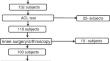
Material and methods: A search was performed to identify meniscal root tears. Age, sex, BMI, and pain were recorded. Knee radiographs and MRI were reviewed. Presence, grade and morphology of MRT, meniscal extrusion, insufficiency fractures, as well as joint structural abnormalities were scored. For goals (I), (II), and (III), tabulations for categorical variables and mean for continuous variables were computed. MRT findings variables were described using percentages. For goal (IV), adjusted linear and logistic regression were employed.
Results: Ninety-six patients with a mean age of 56.6 years (69 females) and mean BMI of 28.9 kg/m2 were included; 88 of the MRT were located at the posterior horn of the medial meniscus (PHMM), and 82% were radial tear. The mean tear diameter was 3.8 mm, and 78/96 tears presented with meniscal extrusion. Nineteen patients presented with subchondral insufficiency fracture (SIF), which was significantly associated with the gap of the tear (p = 0.001) and grade of the meniscal root lesion (p = 0.005).
Conclusion: MRT typically found in middle-aged to older overweight and obese women. Lesions were mostly radial tears and located at PHMM and were frequently associated with meniscal extrusion and SIF. Moreover, the presence of SIF was significantly associated with the gap width and grade of root tear.
期刊介绍:
Skeletal Radiology provides a forum for the dissemination of current knowledge and information dealing with disorders of the musculoskeletal system including the spine. While emphasizing the radiological aspects of the many varied skeletal abnormalities, the journal also adopts an interdisciplinary approach, reflecting the membership of the International Skeletal Society. Thus, the anatomical, pathological, physiological, clinical, metabolic and epidemiological aspects of the many entities affecting the skeleton receive appropriate consideration.
This is the Journal of the International Skeletal Society and the Official Journal of the Society of Skeletal Radiology and the Australasian Musculoskelelal Imaging Group.

 求助内容:
求助内容: 应助结果提醒方式:


