{"title":"Clinical Course of Residual Ventricular Septal Defects After Congenital Heart Disease Repair.","authors":"Yuki Nakayama, Yoshihiko Horimoto, Kazuhiro Suzuki, Makoto Takiguchi, Kazuaki Ishihara, Nobuhiro Umehara, Takeshi Shinkawa","doi":"10.1007/s00246-024-03542-5","DOIUrl":null,"url":null,"abstract":"<p><p>The clinical course of residual ventricular septal defects after congenital heart disease repair is not completely elucidated in the medical literature. This study assessed the incidence, size, and clinical course of residual defects.This single-center retrospective study included 132 patients who survived after ventricular septal defect patch closure (n = 107) and intracardiac repair of double-outlet right ventricle (n = 16) and tetralogy of Fallot (n = 9). Residual defect was evaluated on transthoracic echocardiogram upon hospital discharge and at outpatient clinic visits.The median age at surgery was 1.2 (0.3-13.9) years. In total, 45 (34.1%) patients presented with residual defects upon hospital discharge. The residual defects were within 2 mm (n = 27), 2-3 mm (n = 15), and > 3 mm (n = 3), and the median size was 1.5 (0.5-3.8) mm. There was no late mortality during a median follow-up of 5.4 years. Among 42 residual defects measuring < 3 mm upon hospital discharge, 37 (82.2%) spontaneously closed. Further, five defects decreased in size (1.8 ± 0.6 mm upon hospital discharge vs1.2 ± 0.8 mm at the latest visits, p = 0.15). However, the size of three residual defects measuring > 3 mm upon hospital discharge increased, and two patients required re-surgery for residual defect.Significant residual defect requiring reoperation was rare. In most cases, residual defects measuring < 3 mm upon hospital discharge spontaneously closed within 5 years, and the size of the other defects decreased.</p>","PeriodicalId":19814,"journal":{"name":"Pediatric Cardiology","volume":" ","pages":"1248-1253"},"PeriodicalIF":1.5000,"publicationDate":"2025-06-01","publicationTypes":"Journal Article","fieldsOfStudy":null,"isOpenAccess":false,"openAccessPdf":"","citationCount":"0","resultStr":null,"platform":"Semanticscholar","paperid":null,"PeriodicalName":"Pediatric Cardiology","FirstCategoryId":"3","ListUrlMain":"https://doi.org/10.1007/s00246-024-03542-5","RegionNum":4,"RegionCategory":"医学","ArticlePicture":[],"TitleCN":null,"AbstractTextCN":null,"PMCID":null,"EPubDate":"2024/6/25 0:00:00","PubModel":"Epub","JCR":"Q3","JCRName":"CARDIAC & CARDIOVASCULAR SYSTEMS","Score":null,"Total":0}
引用次数: 0
Abstract
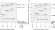
The clinical course of residual ventricular septal defects after congenital heart disease repair is not completely elucidated in the medical literature. This study assessed the incidence, size, and clinical course of residual defects.This single-center retrospective study included 132 patients who survived after ventricular septal defect patch closure (n = 107) and intracardiac repair of double-outlet right ventricle (n = 16) and tetralogy of Fallot (n = 9). Residual defect was evaluated on transthoracic echocardiogram upon hospital discharge and at outpatient clinic visits.The median age at surgery was 1.2 (0.3-13.9) years. In total, 45 (34.1%) patients presented with residual defects upon hospital discharge. The residual defects were within 2 mm (n = 27), 2-3 mm (n = 15), and > 3 mm (n = 3), and the median size was 1.5 (0.5-3.8) mm. There was no late mortality during a median follow-up of 5.4 years. Among 42 residual defects measuring < 3 mm upon hospital discharge, 37 (82.2%) spontaneously closed. Further, five defects decreased in size (1.8 ± 0.6 mm upon hospital discharge vs1.2 ± 0.8 mm at the latest visits, p = 0.15). However, the size of three residual defects measuring > 3 mm upon hospital discharge increased, and two patients required re-surgery for residual defect.Significant residual defect requiring reoperation was rare. In most cases, residual defects measuring < 3 mm upon hospital discharge spontaneously closed within 5 years, and the size of the other defects decreased.
期刊介绍:
The editor of Pediatric Cardiology welcomes original manuscripts concerning all aspects of heart disease in infants, children, and adolescents, including embryology and anatomy, physiology and pharmacology, biochemistry, pathology, genetics, radiology, clinical aspects, investigative cardiology, electrophysiology and echocardiography, and cardiac surgery. Articles which may include original articles, review articles, letters to the editor etc., must be written in English and must be submitted solely to Pediatric Cardiology.
 求助内容:
求助内容: 应助结果提醒方式:


