Juliana Maria Sansevero Senne, Ademir Franco, Carolina de Paula Rossetto Lisboa, José Luiz Cintra Junqueira, Francine Kühl Panzarella, Mariana Quirino Silveira Soares
{"title":"Three-dimensional replica of the temporal bone in the teaching of human anatomy.","authors":"Juliana Maria Sansevero Senne, Ademir Franco, Carolina de Paula Rossetto Lisboa, José Luiz Cintra Junqueira, Francine Kühl Panzarella, Mariana Quirino Silveira Soares","doi":"10.1007/s00276-024-03417-7","DOIUrl":null,"url":null,"abstract":"<p><strong>Purpose: </strong>The current study proposes the comparison of the visualization and identification of anatomical details between natural human temporal bone, its respective copy from three-dimensional printing, and the virtual model obtained from CBCT.</p><p><strong>Methods: </strong>The sample consisted of undergraduate students in Dentistry (Group UE, n = 22), Postgraduate students in Radiology and Imaging (Group P-RI, n = 20), and Postgraduate students in Forensic Odontology (Group P-FO, n = 24). All participants attended a theoretical class on specialized anatomy of the temporal bone and subsequently performed the markings of 10 determined structures.</p><p><strong>Results: </strong>The number of correct identifications was similar in natural bone and printed three-dimensional models in all groups (p > 0.05). The virtual model showed a significantly lower number of correct structures (p < 0.05) in the 3 groups. In general, there were significantly higher percentages of accurate answers among postgraduate students compared to undergraduate students. Most graduate students believed that the printed three-dimensional model could be used to teach anatomy in place of natural bone, while undergraduate students disagreed or were unsure (p < 0.05). Regarding the virtual tomographic image, in all groups, students disagreed or were not sure that its use would be beneficial in replacing natural bone.</p><p><strong>Conclusion: </strong>Three-dimensional and virtual models can be used as auxiliary tools in teaching anatomy, complementing practical learning with natural bones.</p>","PeriodicalId":49461,"journal":{"name":"Surgical and Radiologic Anatomy","volume":" ","pages":"1345-1353"},"PeriodicalIF":1.4000,"publicationDate":"2024-08-01","publicationTypes":"Journal Article","fieldsOfStudy":null,"isOpenAccess":false,"openAccessPdf":"","citationCount":"0","resultStr":null,"platform":"Semanticscholar","paperid":null,"PeriodicalName":"Surgical and Radiologic Anatomy","FirstCategoryId":"3","ListUrlMain":"https://doi.org/10.1007/s00276-024-03417-7","RegionNum":4,"RegionCategory":"医学","ArticlePicture":[],"TitleCN":null,"AbstractTextCN":null,"PMCID":null,"EPubDate":"2024/6/22 0:00:00","PubModel":"Epub","JCR":"Q2","JCRName":"Medicine","Score":null,"Total":0}
引用次数: 0
Abstract
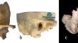
Purpose: The current study proposes the comparison of the visualization and identification of anatomical details between natural human temporal bone, its respective copy from three-dimensional printing, and the virtual model obtained from CBCT.
Methods: The sample consisted of undergraduate students in Dentistry (Group UE, n = 22), Postgraduate students in Radiology and Imaging (Group P-RI, n = 20), and Postgraduate students in Forensic Odontology (Group P-FO, n = 24). All participants attended a theoretical class on specialized anatomy of the temporal bone and subsequently performed the markings of 10 determined structures.
Results: The number of correct identifications was similar in natural bone and printed three-dimensional models in all groups (p > 0.05). The virtual model showed a significantly lower number of correct structures (p < 0.05) in the 3 groups. In general, there were significantly higher percentages of accurate answers among postgraduate students compared to undergraduate students. Most graduate students believed that the printed three-dimensional model could be used to teach anatomy in place of natural bone, while undergraduate students disagreed or were unsure (p < 0.05). Regarding the virtual tomographic image, in all groups, students disagreed or were not sure that its use would be beneficial in replacing natural bone.
Conclusion: Three-dimensional and virtual models can be used as auxiliary tools in teaching anatomy, complementing practical learning with natural bones.
期刊介绍:
Anatomy is a morphological science which cannot fail to interest the clinician. The practical application of anatomical research to clinical problems necessitates special adaptation and selectivity in choosing from numerous international works. Although there is a tendency to believe that meaningful advances in anatomy are unlikely, constant revision is necessary. Surgical and Radiologic Anatomy, the first international journal of Clinical anatomy has been created in this spirit.
Its goal is to serve clinicians, regardless of speciality-physicians, surgeons, radiologists or other specialists-as an indispensable aid with which they can improve their knowledge of anatomy. Each issue includes: Original papers, review articles, articles on the anatomical bases of medical, surgical and radiological techniques, articles of normal radiologic anatomy, brief reviews of anatomical publications of clinical interest.
Particular attention is given to high quality illustrations, which are indispensable for a better understanding of anatomical problems.
Surgical and Radiologic Anatomy is a journal written by anatomists for clinicians with a special interest in anatomy.

 求助内容:
求助内容: 应助结果提醒方式:


