{"title":"Quantitative evaluation of lower limb varicose veins using photoacoustic imaging.","authors":"Moemi Urano, Kenichi Nagae, Sachiko Matsuda, Kentaro Matsubara, Takayuki Yagi, Nobuaki Imanishi, Sadakazu Aiso, Hideaki Obara, Masahiro Jinzaki","doi":"10.1007/s10396-024-01470-8","DOIUrl":null,"url":null,"abstract":"<p><strong>Purpose: </strong>Varicose veins in the lower extremities are dilated subcutaneous varicose veins with a diameter of ≥ 3 mm, caused by increased venous pressure resulting from backflow of blood due to venous valve insufficiency (Gloviczki in Handbook of venous disorders: guidelines of the American venous forum, Hodder Arnold, London, 2009). When diagnosing varicose veins, the shape and thickness of the blood vessels should be accurately visualized in three dimensions. In this study, we investigated a new method for numerical evaluation of vascular morphology related to varicose veins in the lower extremities, using a photoacoustic imaging (PAI) system, which can acquire high-resolution and three-dimensional images noninvasively.</p><p><strong>Methods: </strong>Nine patients with varicose veins participated in the study, and their images were captured using an optical camera and PAI system. We visualized the vascular structure, created a blood presence density (BPD) heat map, and examined the correlation between BPD and location of varicose veins.</p><p><strong>Results: </strong>The obtained photoacoustic (PA) images demonstrated the ability of this method to visualize vessels ranging from as small as 0.2 mm in diameter to large, dilated vessels in three dimensions. Furthermore, the study revealed a correlation between the high-density part of the BPD heat map generated from the PAI images and the presence of varicose veins.</p><p><strong>Conclusion: </strong>PAI is a promising technique for noninvasive and accurate diagnosis of varicose veins in the lower extremities. By providing valuable information on the morphology and hemodynamics of the varicose veins, PAI may facilitate their early detection and treatment.</p>","PeriodicalId":50130,"journal":{"name":"Journal of Medical Ultrasonics","volume":" ","pages":"507-516"},"PeriodicalIF":1.9000,"publicationDate":"2024-07-01","publicationTypes":"Journal Article","fieldsOfStudy":null,"isOpenAccess":false,"openAccessPdf":"https://www.ncbi.nlm.nih.gov/pmc/articles/PMC11272806/pdf/","citationCount":"0","resultStr":null,"platform":"Semanticscholar","paperid":null,"PeriodicalName":"Journal of Medical Ultrasonics","FirstCategoryId":"3","ListUrlMain":"https://doi.org/10.1007/s10396-024-01470-8","RegionNum":4,"RegionCategory":"医学","ArticlePicture":[],"TitleCN":null,"AbstractTextCN":null,"PMCID":null,"EPubDate":"2024/6/20 0:00:00","PubModel":"Epub","JCR":"Q3","JCRName":"RADIOLOGY, NUCLEAR MEDICINE & MEDICAL IMAGING","Score":null,"Total":0}
引用次数: 0
Abstract
Purpose: Varicose veins in the lower extremities are dilated subcutaneous varicose veins with a diameter of ≥ 3 mm, caused by increased venous pressure resulting from backflow of blood due to venous valve insufficiency (Gloviczki in Handbook of venous disorders: guidelines of the American venous forum, Hodder Arnold, London, 2009). When diagnosing varicose veins, the shape and thickness of the blood vessels should be accurately visualized in three dimensions. In this study, we investigated a new method for numerical evaluation of vascular morphology related to varicose veins in the lower extremities, using a photoacoustic imaging (PAI) system, which can acquire high-resolution and three-dimensional images noninvasively.
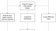
Methods: Nine patients with varicose veins participated in the study, and their images were captured using an optical camera and PAI system. We visualized the vascular structure, created a blood presence density (BPD) heat map, and examined the correlation between BPD and location of varicose veins.
Results: The obtained photoacoustic (PA) images demonstrated the ability of this method to visualize vessels ranging from as small as 0.2 mm in diameter to large, dilated vessels in three dimensions. Furthermore, the study revealed a correlation between the high-density part of the BPD heat map generated from the PAI images and the presence of varicose veins.
Conclusion: PAI is a promising technique for noninvasive and accurate diagnosis of varicose veins in the lower extremities. By providing valuable information on the morphology and hemodynamics of the varicose veins, PAI may facilitate their early detection and treatment.
期刊介绍:
The Journal of Medical Ultrasonics is the official journal of the Japan Society of Ultrasonics in Medicine. The main purpose of the journal is to provide forum for the publication of papers documenting recent advances and new developments in the entire field of ultrasound in medicine and biology, encompassing both the medical and the engineering aspects of the science.The journal welcomes original articles, review articles, images, and letters to the editor.The journal also provides state-of-the-art information such as announcements from the boards and the committees of the society.
 求助内容:
求助内容: 应助结果提醒方式:


