Joao G Porto, Ruben Blachman-Braun, Archan Khandekar, Oleksandr N Kryvenko, Dipen J Parekh
{"title":"Chondroma in the Urinary Bladder: An Extremely Rare Finding.","authors":"Joao G Porto, Ruben Blachman-Braun, Archan Khandekar, Oleksandr N Kryvenko, Dipen J Parekh","doi":"10.1155/2024/4120514","DOIUrl":null,"url":null,"abstract":"<p><p>Chondroma, commonly observed in the bones, has limited documentation when found in soft tissues. To date, only 8 chondromas in the urinary bladder have been reported, all in females. Here, we describe a 54-year-old female who presented with a chondroma located at the anterior wall of the urinary bladder. An incidental 5 mm enhanced focus was identified on the right bladder wall during a contrast-enhanced computerized tomography (CT). Subsequent cystoscopy did not reveal any abnormalities, and both urinalysis and urine cytology were unremarkable. However, a CT urogram reconfirmed suspicions of malignancy, which a cystoscopy validated. The patient underwent a transurethral resection of the bladder tumor, which was identified as a bladder chondroma. During the surgical incision, a submucosal lesion was found, which was further confirmed with histopathological evaluation. Over a year-long follow-up using imaging and urine cytology, no recurrence was observed. This case reinforces earlier findings and underscores the predilection for females between their 5<sup>th</sup> and 7<sup>th</sup> decades with a positive prognosis.</p>","PeriodicalId":30323,"journal":{"name":"Case Reports in Urology","volume":"2024 ","pages":"4120514"},"PeriodicalIF":0.0000,"publicationDate":"2024-06-06","publicationTypes":"Journal Article","fieldsOfStudy":null,"isOpenAccess":false,"openAccessPdf":"https://www.ncbi.nlm.nih.gov/pmc/articles/PMC11178419/pdf/","citationCount":"0","resultStr":null,"platform":"Semanticscholar","paperid":null,"PeriodicalName":"Case Reports in Urology","FirstCategoryId":"1085","ListUrlMain":"https://doi.org/10.1155/2024/4120514","RegionNum":0,"RegionCategory":null,"ArticlePicture":[],"TitleCN":null,"AbstractTextCN":null,"PMCID":null,"EPubDate":"2024/1/1 0:00:00","PubModel":"eCollection","JCR":"","JCRName":"","Score":null,"Total":0}
引用次数: 0
Abstract
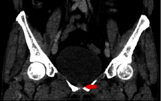
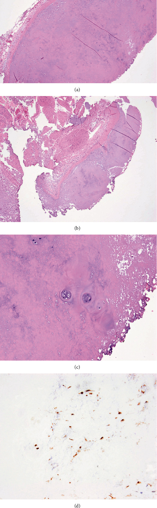
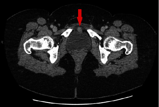
Chondroma, commonly observed in the bones, has limited documentation when found in soft tissues. To date, only 8 chondromas in the urinary bladder have been reported, all in females. Here, we describe a 54-year-old female who presented with a chondroma located at the anterior wall of the urinary bladder. An incidental 5 mm enhanced focus was identified on the right bladder wall during a contrast-enhanced computerized tomography (CT). Subsequent cystoscopy did not reveal any abnormalities, and both urinalysis and urine cytology were unremarkable. However, a CT urogram reconfirmed suspicions of malignancy, which a cystoscopy validated. The patient underwent a transurethral resection of the bladder tumor, which was identified as a bladder chondroma. During the surgical incision, a submucosal lesion was found, which was further confirmed with histopathological evaluation. Over a year-long follow-up using imaging and urine cytology, no recurrence was observed. This case reinforces earlier findings and underscores the predilection for females between their 5th and 7th decades with a positive prognosis.

 求助内容:
求助内容: 应助结果提醒方式:


